
While the vast majority of cancer diagnoses are made or confirmed by a pathologist, the increased demand for pathology services is not being matched by a growing number of skilled pathologists. The largest pathology laboratory in the Netherlands, LabPON, tells how digitizing their pathology workflow with the Philips IntelliSite is helping them to advance its services. This material is not for distribution/use in the USA
The clinical need
U.S. figures show that the the number of active physicians in pathology decreased by 7.4% between 2000 and 2010, and 57.3% of active physicians in pathology are 55 or older.¹ At the same time, there is room to improve the accuracy of diagnosis and the speed of pathology services. For example, one article reported that approximately 20% of current HER2 testing may be inaccurate.² Another report said that pathology accounts for 41% of delays in cancer diagnosis, and is a leading cause of patient safety incidents.³
Speeding results for 90,000 pathology cases a year
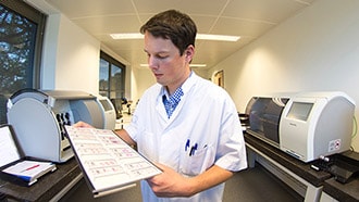
Handling over ninety thousand cases per year, LabPON knows that patients rely on them….and waiting for the lab results can cause fear and anxiety. LabPON chose the Philips IntelliSite pathology solution to gradually move to a complete digital workflow. It digitizes the images that pathologists normally view through a microscope using an advanced slide scanner and image management system. This is helping them improve the speed and quality of their services.
“Since the files are digital, a colleague can access them digitally regardless of a location. You no longer need to worry about the difficult logistics involved in sending glass slides. You no longer run the risk of losing or damaging specimens. You save an enormous amount of money, and you save a lot of time in the consulting process.”
Alexi Baidoshvili
MD, Pathologist, LabPON
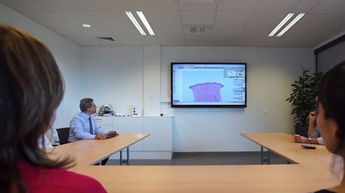
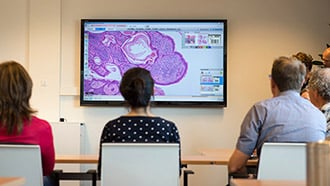
Watch video about how LabPON has improved their pathology services.
How does it work
The Philips IntelliSite pathology solution consists of an advanced slide scanner and image management system. To support high-volume pathology labs, the IntelliSite Ultra-Fast Scanner (UFS) scans all slides at high resolution, providing visibility to cellular detail. A single slide can be scanned in 60 seconds at 40x equivalent for 15mm x 15mm scan area. The IntelliSite Image Management System (IMS) allows you to process, store, view and share images across your entire enterprise. Philips Digital Pathology Solutions has acquired the CE mark for diagnosis of routine pathology, including Hematoxylin and Eosin (H&E), Immunohistochemistry (IHC), and special stained formalin-fixed, paraffin-embedded tissue sections.
For more information
Written by:

Hans Driessen Senior Communications Manager, Philips Digital Pathology Solutions
Share this article
Related articles


¹⋅AAMC 2012 Physician Specialty Data Book - Center for Workforce Studies November 2012 ²⋅Wolff AC et al. American Society of Clinical Oncology/College of American Pathologists guideline recommendations for human epidermal growth factor receptor 2 testing in breast cancer. J Clinical Oncology (2007) 25: 118-145. ³⋅National Painet Safety Agency - Delayed diagnosis of cancer Thematic review - National Patient Safety Agency, March 2010