VesselNavigator

3D image fusion technology for endovascular procedures

Find similar products

VesselNavigator allows reuse of 3D vascular anatomical information from existing CTA and MRA datasets as a 3D roadmap overlay on a live X-ray image. With its excellent visualization, VesselNavigator provides an intuitive and continuous 3D roadmap to guide you through vasculature during the entire procedure.

Features
3D image fusion
3D image fusion in endovascular procedures
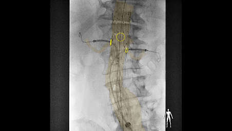
3D image fusion in endovascular procedures

Studies¹,² have shown the advantages of a 3D perspective for catheter and device guidance. With VesselNavigator, clinicians can easily segment 3D vessel structures from existing CT/MR datasets and fuse them with live X-ray for guidance.

3D image fusion in endovascular procedures

3D image fusion in endovascular procedures
Studies¹,² have shown the advantages of a 3D perspective for catheter and device guidance. With VesselNavigator, clinicians can easily segment 3D vessel structures from existing CT/MR datasets and fuse them with live X-ray for guidance.

3D image fusion in endovascular procedures

Studies¹,² have shown the advantages of a 3D perspective for catheter and device guidance. With VesselNavigator, clinicians can easily segment 3D vessel structures from existing CT/MR datasets and fuse them with live X-ray for guidance.
Click here for more information
3D image fusion
3D image fusion in endovascular procedures

3D image fusion in endovascular procedures

Studies¹,² have shown the advantages of a 3D perspective for catheter and device guidance. With VesselNavigator, clinicians can easily segment 3D vessel structures from existing CT/MR datasets and fuse them with live X-ray for guidance.
Reduction in contrast usage
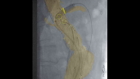
Reduction in contrast usage

Average contrast medium usage was reduced by 72% in a study¹ that used Philips CTA image fusion guidance during endovascular repair of complex aortic aneurysms. No intraprocedural contrast agent injection was required to create a roadmap.

Reduction in contrast usage

Reduction in contrast usage
Average contrast medium usage was reduced by 72% in a study¹ that used Philips CTA image fusion guidance during endovascular repair of complex aortic aneurysms. No intraprocedural contrast agent injection was required to create a roadmap.

Reduction in contrast usage

Average contrast medium usage was reduced by 72% in a study¹ that used Philips CTA image fusion guidance during endovascular repair of complex aortic aneurysms. No intraprocedural contrast agent injection was required to create a roadmap.
Click here for more information
Reduction in contrast usage

Reduction in contrast usage

Average contrast medium usage was reduced by 72% in a study¹ that used Philips CTA image fusion guidance during endovascular repair of complex aortic aneurysms. No intraprocedural contrast agent injection was required to create a roadmap.
Potential to reduce procedure time
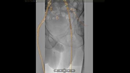
Potential to reduce procedure time

Philips CTA Image Fusion Guidance may lead to shorter procedure times. A study of 62 patients² showed an average reduction in procedure time from 6.3 to 5.2 hours during FEVAR/BEVAR procedures using Philips CTA image fusion guidance.

Potential to reduce procedure time

Potential to reduce procedure time
Philips CTA Image Fusion Guidance may lead to shorter procedure times. A study of 62 patients² showed an average reduction in procedure time from 6.3 to 5.2 hours during FEVAR/BEVAR procedures using Philips CTA image fusion guidance.

Potential to reduce procedure time

Philips CTA Image Fusion Guidance may lead to shorter procedure times. A study of 62 patients² showed an average reduction in procedure time from 6.3 to 5.2 hours during FEVAR/BEVAR procedures using Philips CTA image fusion guidance.
Click here for more information
Potential to reduce procedure time

Potential to reduce procedure time

Philips CTA Image Fusion Guidance may lead to shorter procedure times. A study of 62 patients² showed an average reduction in procedure time from 6.3 to 5.2 hours during FEVAR/BEVAR procedures using Philips CTA image fusion guidance.
Moves in sync to new positions
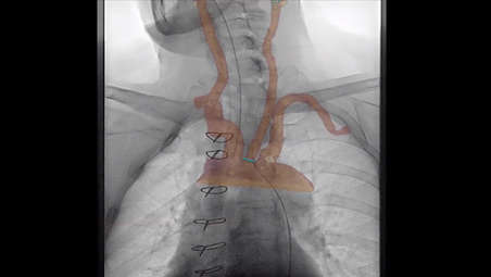
Moves in sync to new positions

During the procedure, VesselNavigator provides a real-time overlay that moves in sync with any projection, table and system position. This can reduce the need to make additional contrast enhanced runs to create new roadmaps.

Moves in sync to new positions

Moves in sync to new positions
During the procedure, VesselNavigator provides a real-time overlay that moves in sync with any projection, table and system position. This can reduce the need to make additional contrast enhanced runs to create new roadmaps.

Moves in sync to new positions

During the procedure, VesselNavigator provides a real-time overlay that moves in sync with any projection, table and system position. This can reduce the need to make additional contrast enhanced runs to create new roadmaps.
Click here for more information
Moves in sync to new positions

Moves in sync to new positions

During the procedure, VesselNavigator provides a real-time overlay that moves in sync with any projection, table and system position. This can reduce the need to make additional contrast enhanced runs to create new roadmaps.
Superb image quality

Superb image quality

Create overlays based on a variety of superb volume rendering visualization options. These can be customized according to user preferences.

Superb image quality

Superb image quality
Create overlays based on a variety of superb volume rendering visualization options. These can be customized according to user preferences.

Superb image quality

Create overlays based on a variety of superb volume rendering visualization options. These can be customized according to user preferences.
Click here for more information
Superb image quality

Superb image quality

Create overlays based on a variety of superb volume rendering visualization options. These can be customized according to user preferences.
Anatomical
Anatomical ring markers

Anatomical ring markers
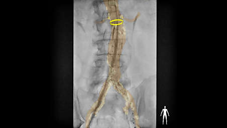
Ring markers can be added to the segmented image to indicate the ostia and landing zone and to define planning angles. These markers are visualized during the procedure for guidance.

Anatomical ring markers

Anatomical ring markers
Ring markers can be added to the segmented image to indicate the ostia and landing zone and to define planning angles. These markers are visualized during the procedure for guidance.

Anatomical ring markers

Ring markers can be added to the segmented image to indicate the ostia and landing zone and to define planning angles. These markers are visualized during the procedure for guidance.
Click here for more information
Anatomical
Anatomical ring markers

Anatomical ring markers

Ring markers can be added to the segmented image to indicate the ostia and landing zone and to define planning angles. These markers are visualized during the procedure for guidance.
  • 3D image fusion
  • Reduction in contrast usage
  • Potential to reduce procedure time
  • Moves in sync to new positions
See all features
3D image fusion
3D image fusion in endovascular procedures

3D image fusion in endovascular procedures

Studies¹,² have shown the advantages of a 3D perspective for catheter and device guidance. With VesselNavigator, clinicians can easily segment 3D vessel structures from existing CT/MR datasets and fuse them with live X-ray for guidance.

3D image fusion in endovascular procedures

3D image fusion in endovascular procedures
Studies¹,² have shown the advantages of a 3D perspective for catheter and device guidance. With VesselNavigator, clinicians can easily segment 3D vessel structures from existing CT/MR datasets and fuse them with live X-ray for guidance.

3D image fusion in endovascular procedures

Studies¹,² have shown the advantages of a 3D perspective for catheter and device guidance. With VesselNavigator, clinicians can easily segment 3D vessel structures from existing CT/MR datasets and fuse them with live X-ray for guidance.
Click here for more information
3D image fusion
3D image fusion in endovascular procedures

3D image fusion in endovascular procedures

Studies¹,² have shown the advantages of a 3D perspective for catheter and device guidance. With VesselNavigator, clinicians can easily segment 3D vessel structures from existing CT/MR datasets and fuse them with live X-ray for guidance.
Reduction in contrast usage

Reduction in contrast usage

Average contrast medium usage was reduced by 72% in a study¹ that used Philips CTA image fusion guidance during endovascular repair of complex aortic aneurysms. No intraprocedural contrast agent injection was required to create a roadmap.

Reduction in contrast usage

Reduction in contrast usage
Average contrast medium usage was reduced by 72% in a study¹ that used Philips CTA image fusion guidance during endovascular repair of complex aortic aneurysms. No intraprocedural contrast agent injection was required to create a roadmap.

Reduction in contrast usage

Average contrast medium usage was reduced by 72% in a study¹ that used Philips CTA image fusion guidance during endovascular repair of complex aortic aneurysms. No intraprocedural contrast agent injection was required to create a roadmap.
Click here for more information
Reduction in contrast usage

Reduction in contrast usage

Average contrast medium usage was reduced by 72% in a study¹ that used Philips CTA image fusion guidance during endovascular repair of complex aortic aneurysms. No intraprocedural contrast agent injection was required to create a roadmap.
Potential to reduce procedure time

Potential to reduce procedure time

Philips CTA Image Fusion Guidance may lead to shorter procedure times. A study of 62 patients² showed an average reduction in procedure time from 6.3 to 5.2 hours during FEVAR/BEVAR procedures using Philips CTA image fusion guidance.

Potential to reduce procedure time

Potential to reduce procedure time
Philips CTA Image Fusion Guidance may lead to shorter procedure times. A study of 62 patients² showed an average reduction in procedure time from 6.3 to 5.2 hours during FEVAR/BEVAR procedures using Philips CTA image fusion guidance.

Potential to reduce procedure time

Philips CTA Image Fusion Guidance may lead to shorter procedure times. A study of 62 patients² showed an average reduction in procedure time from 6.3 to 5.2 hours during FEVAR/BEVAR procedures using Philips CTA image fusion guidance.
Click here for more information
Potential to reduce procedure time

Potential to reduce procedure time

Philips CTA Image Fusion Guidance may lead to shorter procedure times. A study of 62 patients² showed an average reduction in procedure time from 6.3 to 5.2 hours during FEVAR/BEVAR procedures using Philips CTA image fusion guidance.
Moves in sync to new positions

Moves in sync to new positions

During the procedure, VesselNavigator provides a real-time overlay that moves in sync with any projection, table and system position. This can reduce the need to make additional contrast enhanced runs to create new roadmaps.

Moves in sync to new positions

Moves in sync to new positions
During the procedure, VesselNavigator provides a real-time overlay that moves in sync with any projection, table and system position. This can reduce the need to make additional contrast enhanced runs to create new roadmaps.

Moves in sync to new positions

During the procedure, VesselNavigator provides a real-time overlay that moves in sync with any projection, table and system position. This can reduce the need to make additional contrast enhanced runs to create new roadmaps.
Click here for more information
Moves in sync to new positions

Moves in sync to new positions

During the procedure, VesselNavigator provides a real-time overlay that moves in sync with any projection, table and system position. This can reduce the need to make additional contrast enhanced runs to create new roadmaps.
Superb image quality

Superb image quality

Create overlays based on a variety of superb volume rendering visualization options. These can be customized according to user preferences.

Superb image quality

Superb image quality
Create overlays based on a variety of superb volume rendering visualization options. These can be customized according to user preferences.

Superb image quality

Create overlays based on a variety of superb volume rendering visualization options. These can be customized according to user preferences.
Click here for more information
Superb image quality

Superb image quality

Create overlays based on a variety of superb volume rendering visualization options. These can be customized according to user preferences.
Anatomical
Anatomical ring markers

Anatomical ring markers

Ring markers can be added to the segmented image to indicate the ostia and landing zone and to define planning angles. These markers are visualized during the procedure for guidance.

Anatomical ring markers

Anatomical ring markers
Ring markers can be added to the segmented image to indicate the ostia and landing zone and to define planning angles. These markers are visualized during the procedure for guidance.

Anatomical ring markers

Ring markers can be added to the segmented image to indicate the ostia and landing zone and to define planning angles. These markers are visualized during the procedure for guidance.
Click here for more information
Anatomical
Anatomical ring markers

Anatomical ring markers

Ring markers can be added to the segmented image to indicate the ostia and landing zone and to define planning angles. These markers are visualized during the procedure for guidance.
  • 1. Tacher V, et al (2013). Image Guidance for Endovascular Repair of Complex Aortic Aneurysms: Comparison of Two-dimensional and Three-dimensional Angiography and Image Fusion, J Vasc Interv Radiol, 24(11), 1698-1706. http://www.ncbi.nlm.nih.gov/pubmed/24035418
  • 2. Sailer AM, et al (2014). CTA with fluoroscopy image fusion guidance in endovascular complex aortic aneurysm repair, Eur J Vasc Endovasc Surg. 2014 Apr;47(4):349-56. http://www.ncbi.nlm.nih.gov/pubmed/24485850
You are about to visit a Philips global content page

Continue

You are about to visit a Philips global content page

Continue

Our site can best be viewed with the latest version of Microsoft Edge, Google Chrome or Firefox.

Jūs ieejat Latvijas Philips Healthcare tīmekļa vietnē.
Please select the checkbox

Tīmekļa vietne ir paredzēta tikai personām, kuras izmanto medicīniskās ierīces kā profesionāļi, tostarp personām, kas praktizē medicīnas profesijās, personām, kas darbojas veselības aprūpes iestāžu labā vai organizācijām, kas tirgo medicīnas ierīces kā profesionāļi.

Vai esat persona, kas atbilst iepriekš minētajiem kritērijiem?