Affiniti 70

Ultrasound system

Find similar products

Philips understand the challenges you face. From a changing patient population to the need to maximize resources, the challenges can be considerable. Philips Affiniti helps you overcome these daily challenges so you can provide the best possible care for your patients every day. Affiniti offers a powerful combination of performance and workflow advances to enhance diagnostic confidence making it the choice of clinicians worldwide.

Features
Next Gen AutoSCAN
Next Gen AutoSCAN

Next Gen AutoSCAN

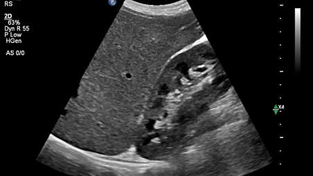
Philips Next Gen AutoSCAN improves image uniformity, adaptively adjusting image brightness at every pixel and reducing the need for user adjustment while also improving transducer plunkability. Next Gen AutoSCAN reduces button pushes by up to 54% with pixel-by-pixel real-time optimization.

Next Gen AutoSCAN

Next Gen AutoSCAN
Philips Next Gen AutoSCAN improves image uniformity, adaptively adjusting image brightness at every pixel and reducing the need for user adjustment while also improving transducer plunkability. Next Gen AutoSCAN reduces button pushes by up to 54% with pixel-by-pixel real-time optimization.

Next Gen AutoSCAN

Philips Next Gen AutoSCAN improves image uniformity, adaptively adjusting image brightness at every pixel and reducing the need for user adjustment while also improving transducer plunkability. Next Gen AutoSCAN reduces button pushes by up to 54% with pixel-by-pixel real-time optimization.
Click here for more information
Next Gen AutoSCAN
Next Gen AutoSCAN

Next Gen AutoSCAN

Philips Next Gen AutoSCAN improves image uniformity, adaptively adjusting image brightness at every pixel and reducing the need for user adjustment while also improving transducer plunkability. Next Gen AutoSCAN reduces button pushes by up to 54% with pixel-by-pixel real-time optimization.
Flow Viewer
Flow Viewer

Flow Viewer

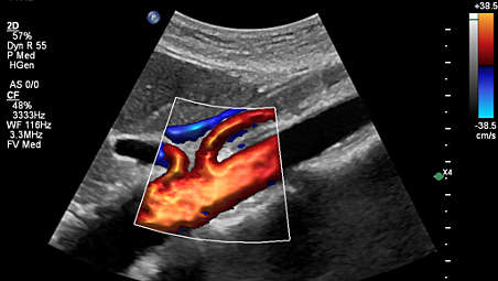
Flow Viewer is a Philips color visualization enhancement for vasculature and fetal heart architecture. Flow Viewer provides a 3D-like rendering of flow imaging data to better visualize the cardiac and vascular architecture and enhance the aesthetic appeal of all color imaging modes Available in all color imaging modes (CFM, CPA, CPAd, MFI, MFI HD).

Flow Viewer

Flow Viewer
Flow Viewer is a Philips color visualization enhancement for vasculature and fetal heart architecture. Flow Viewer provides a 3D-like rendering of flow imaging data to better visualize the cardiac and vascular architecture and enhance the aesthetic appeal of all color imaging modes Available in all color imaging modes (CFM, CPA, CPAd, MFI, MFI HD).

Flow Viewer

Flow Viewer is a Philips color visualization enhancement for vasculature and fetal heart architecture. Flow Viewer provides a 3D-like rendering of flow imaging data to better visualize the cardiac and vascular architecture and enhance the aesthetic appeal of all color imaging modes Available in all color imaging modes (CFM, CPA, CPAd, MFI, MFI HD).
Click here for more information
Flow Viewer
Flow Viewer

Flow Viewer

Flow Viewer is a Philips color visualization enhancement for vasculature and fetal heart architecture. Flow Viewer provides a 3D-like rendering of flow imaging data to better visualize the cardiac and vascular architecture and enhance the aesthetic appeal of all color imaging modes Available in all color imaging modes (CFM, CPA, CPAd, MFI, MFI HD).
MFI
MFI

MFI

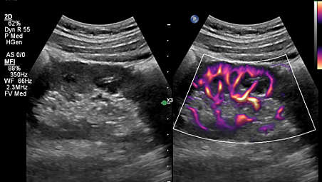
Designed to detect slow and weak blood flow anatomy in tissue. This proprietary approach overcomes many of the barriers associated with conventional methods to detect small vessel blood flow with high resolution and minimal artifacts. MicroFlow Imaging maintains high frame rate and 2D image quality while applying advanced artifact reduction techniques to reveal small vessel anatomy

MFI

MFI
Designed to detect slow and weak blood flow anatomy in tissue. This proprietary approach overcomes many of the barriers associated with conventional methods to detect small vessel blood flow with high resolution and minimal artifacts. MicroFlow Imaging maintains high frame rate and 2D image quality while applying advanced artifact reduction techniques to reveal small vessel anatomy

MFI

Designed to detect slow and weak blood flow anatomy in tissue. This proprietary approach overcomes many of the barriers associated with conventional methods to detect small vessel blood flow with high resolution and minimal artifacts. MicroFlow Imaging maintains high frame rate and 2D image quality while applying advanced artifact reduction techniques to reveal small vessel anatomy
Click here for more information
MFI
MFI

MFI

Designed to detect slow and weak blood flow anatomy in tissue. This proprietary approach overcomes many of the barriers associated with conventional methods to detect small vessel blood flow with high resolution and minimal artifacts. MicroFlow Imaging maintains high frame rate and 2D image quality while applying advanced artifact reduction techniques to reveal small vessel anatomy
Collaboration Live
Collaboration Live
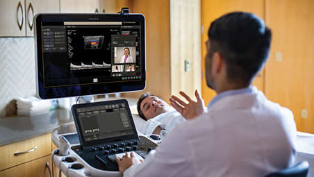
Collaboration Live

Extend your team without expanding it. Collaboration Live is a communication platform that faciitates communication between a compatible ultrasound system and a remote user. With simultaneous Multi-party communication up to six users can quickly and securely talk, text, screen share and video stream direcly from the ultrasound system for access to mulitple clinical resources at a distance.

Collaboration Live

Collaboration Live
Extend your team without expanding it. Collaboration Live is a communication platform that faciitates communication between a compatible ultrasound system and a remote user. With simultaneous Multi-party communication up to six users can quickly and securely talk, text, screen share and video stream direcly from the ultrasound system for access to mulitple clinical resources at a distance.

Collaboration Live

Extend your team without expanding it. Collaboration Live is a communication platform that faciitates communication between a compatible ultrasound system and a remote user. With simultaneous Multi-party communication up to six users can quickly and securely talk, text, screen share and video stream direcly from the ultrasound system for access to mulitple clinical resources at a distance.
Click here for more information
Collaboration Live
Collaboration Live

Collaboration Live

Extend your team without expanding it. Collaboration Live is a communication platform that faciitates communication between a compatible ultrasound system and a remote user. With simultaneous Multi-party communication up to six users can quickly and securely talk, text, screen share and video stream direcly from the ultrasound system for access to mulitple clinical resources at a distance.
Fusion and Navigation
Fusion and Navigation

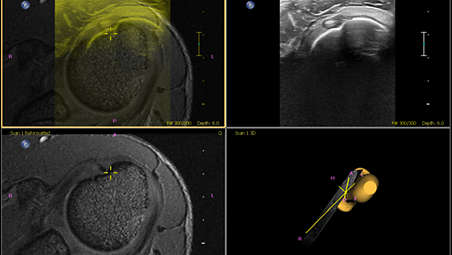
Make confident decisions even in challenging diagnostic cases with fully integrated fusion capabilities that feature streamlined workflows to allow clinicians to achieve fast and effective fusion of CT/MR/PET with live ultrasound. By combining imaging modalities directly on the ultrasound system, you now have access to an even more powerful diagnostic tool with advanced visualization allowing for fast clinical decisions. Expand fusion and navigation capabilities through a range of transducers across applications, including the X6-1 xMatrix , C5-1, C9-2, eL18-4, L12-5, C10-4ec, S5-1 and the new mC7-2

Fusion and Navigation

Fusion and Navigation
Make confident decisions even in challenging diagnostic cases with fully integrated fusion capabilities that feature streamlined workflows to allow clinicians to achieve fast and effective fusion of CT/MR/PET with live ultrasound. By combining imaging modalities directly on the ultrasound system, you now have access to an even more powerful diagnostic tool with advanced visualization allowing for fast clinical decisions. Expand fusion and navigation capabilities through a range of transducers across applications, including the X6-1 xMatrix , C5-1, C9-2, eL18-4, L12-5, C10-4ec, S5-1 and the new mC7-2

Fusion and Navigation

Make confident decisions even in challenging diagnostic cases with fully integrated fusion capabilities that feature streamlined workflows to allow clinicians to achieve fast and effective fusion of CT/MR/PET with live ultrasound. By combining imaging modalities directly on the ultrasound system, you now have access to an even more powerful diagnostic tool with advanced visualization allowing for fast clinical decisions. Expand fusion and navigation capabilities through a range of transducers across applications, including the X6-1 xMatrix , C5-1, C9-2, eL18-4, L12-5, C10-4ec, S5-1 and the new mC7-2
Click here for more information
Fusion and Navigation

Fusion and Navigation

Make confident decisions even in challenging diagnostic cases with fully integrated fusion capabilities that feature streamlined workflows to allow clinicians to achieve fast and effective fusion of CT/MR/PET with live ultrasound. By combining imaging modalities directly on the ultrasound system, you now have access to an even more powerful diagnostic tool with advanced visualization allowing for fast clinical decisions. Expand fusion and navigation capabilities through a range of transducers across applications, including the X6-1 xMatrix , C5-1, C9-2, eL18-4, L12-5, C10-4ec, S5-1 and the new mC7-2
FlexVue with Orthogonal View
FlexVue with Orthogonal View

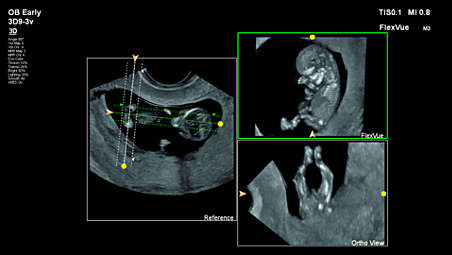
95% of OB/GYN users surveyed* feel, FlexVue and Orthogonal View, improves their workflow and 85% say it enhances their diagnostic confidence as well.

FlexVue with Orthogonal View

FlexVue with Orthogonal View
95% of OB/GYN users surveyed* feel, FlexVue and Orthogonal View, improves their workflow and 85% say it enhances their diagnostic confidence as well.

FlexVue with Orthogonal View

95% of OB/GYN users surveyed* feel, FlexVue and Orthogonal View, improves their workflow and 85% say it enhances their diagnostic confidence as well.
Click here for more information
FlexVue with Orthogonal View

FlexVue with Orthogonal View

95% of OB/GYN users surveyed* feel, FlexVue and Orthogonal View, improves their workflow and 85% say it enhances their diagnostic confidence as well.
PureWave & xMatrix technology
PureWave & xMatrix transducer technology

PureWave & xMatrix transducer technology

The power of PureWave for exceptional imaging even on technically difficult patients. PureWave crystal technology represents the biggest breakthrough in piezoelectric transducer material in 40 years. The pure, uniform crystals of PureWave have virtually perfect uniformity for greater bandwidth and twice the efficiency of conventional ceramic materials.

PureWave & xMatrix transducer technology

PureWave & xMatrix transducer technology
The power of PureWave for exceptional imaging even on technically difficult patients. PureWave crystal technology represents the biggest breakthrough in piezoelectric transducer material in 40 years. The pure, uniform crystals of PureWave have virtually perfect uniformity for greater bandwidth and twice the efficiency of conventional ceramic materials.

PureWave & xMatrix transducer technology

The power of PureWave for exceptional imaging even on technically difficult patients. PureWave crystal technology represents the biggest breakthrough in piezoelectric transducer material in 40 years. The pure, uniform crystals of PureWave have virtually perfect uniformity for greater bandwidth and twice the efficiency of conventional ceramic materials.
Click here for more information
PureWave & xMatrix technology
PureWave & xMatrix transducer technology

PureWave & xMatrix transducer technology

The power of PureWave for exceptional imaging even on technically difficult patients. PureWave crystal technology represents the biggest breakthrough in piezoelectric transducer material in 40 years. The pure, uniform crystals of PureWave have virtually perfect uniformity for greater bandwidth and twice the efficiency of conventional ceramic materials.
TrueVue
TrueVue advanced 3D display

TrueVue advanced 3D display
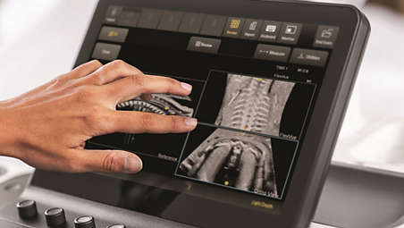
Philips TrueVue advanced 3D ultrasound display delivers amazing lifelike fetal 3D images. TrueVue, with its internal light source, gives clinicians the ability to manipulate light and shadow anywhere in the 3D volume.

TrueVue advanced 3D display

TrueVue advanced 3D display
Philips TrueVue advanced 3D ultrasound display delivers amazing lifelike fetal 3D images. TrueVue, with its internal light source, gives clinicians the ability to manipulate light and shadow anywhere in the 3D volume.

TrueVue advanced 3D display

Philips TrueVue advanced 3D ultrasound display delivers amazing lifelike fetal 3D images. TrueVue, with its internal light source, gives clinicians the ability to manipulate light and shadow anywhere in the 3D volume.
Click here for more information
TrueVue
TrueVue advanced 3D display

TrueVue advanced 3D display

Philips TrueVue advanced 3D ultrasound display delivers amazing lifelike fetal 3D images. TrueVue, with its internal light source, gives clinicians the ability to manipulate light and shadow anywhere in the 3D volume.
MaxVue high definition display
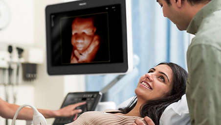
MaxVue high definition display

At the touch of a button, MaxVue high-definition display brings extraordinary visualization of anatomy with 1,179,648 additional image pixels compared to a standard 4:3 display format mode. MaxVue enhances ultrasound viewing and provides 38% more viewing area to optimize the display of dual, side/side, biplane, and scrolling imaging modes.

MaxVue high definition display

MaxVue high definition display
At the touch of a button, MaxVue high-definition display brings extraordinary visualization of anatomy with 1,179,648 additional image pixels compared to a standard 4:3 display format mode. MaxVue enhances ultrasound viewing and provides 38% more viewing area to optimize the display of dual, side/side, biplane, and scrolling imaging modes.

MaxVue high definition display

At the touch of a button, MaxVue high-definition display brings extraordinary visualization of anatomy with 1,179,648 additional image pixels compared to a standard 4:3 display format mode. MaxVue enhances ultrasound viewing and provides 38% more viewing area to optimize the display of dual, side/side, biplane, and scrolling imaging modes.
Click here for more information
MaxVue high definition display

MaxVue high definition display

At the touch of a button, MaxVue high-definition display brings extraordinary visualization of anatomy with 1,179,648 additional image pixels compared to a standard 4:3 display format mode. MaxVue enhances ultrasound viewing and provides 38% more viewing area to optimize the display of dual, side/side, biplane, and scrolling imaging modes.
  • Next Gen AutoSCAN
  • Flow Viewer
  • MFI
  • Collaboration Live
See all features
Next Gen AutoSCAN
Next Gen AutoSCAN

Next Gen AutoSCAN

Philips Next Gen AutoSCAN improves image uniformity, adaptively adjusting image brightness at every pixel and reducing the need for user adjustment while also improving transducer plunkability. Next Gen AutoSCAN reduces button pushes by up to 54% with pixel-by-pixel real-time optimization.

Next Gen AutoSCAN

Next Gen AutoSCAN
Philips Next Gen AutoSCAN improves image uniformity, adaptively adjusting image brightness at every pixel and reducing the need for user adjustment while also improving transducer plunkability. Next Gen AutoSCAN reduces button pushes by up to 54% with pixel-by-pixel real-time optimization.

Next Gen AutoSCAN

Philips Next Gen AutoSCAN improves image uniformity, adaptively adjusting image brightness at every pixel and reducing the need for user adjustment while also improving transducer plunkability. Next Gen AutoSCAN reduces button pushes by up to 54% with pixel-by-pixel real-time optimization.
Click here for more information
Next Gen AutoSCAN
Next Gen AutoSCAN

Next Gen AutoSCAN

Philips Next Gen AutoSCAN improves image uniformity, adaptively adjusting image brightness at every pixel and reducing the need for user adjustment while also improving transducer plunkability. Next Gen AutoSCAN reduces button pushes by up to 54% with pixel-by-pixel real-time optimization.
Flow Viewer
Flow Viewer

Flow Viewer

Flow Viewer is a Philips color visualization enhancement for vasculature and fetal heart architecture. Flow Viewer provides a 3D-like rendering of flow imaging data to better visualize the cardiac and vascular architecture and enhance the aesthetic appeal of all color imaging modes Available in all color imaging modes (CFM, CPA, CPAd, MFI, MFI HD).

Flow Viewer

Flow Viewer
Flow Viewer is a Philips color visualization enhancement for vasculature and fetal heart architecture. Flow Viewer provides a 3D-like rendering of flow imaging data to better visualize the cardiac and vascular architecture and enhance the aesthetic appeal of all color imaging modes Available in all color imaging modes (CFM, CPA, CPAd, MFI, MFI HD).

Flow Viewer

Flow Viewer is a Philips color visualization enhancement for vasculature and fetal heart architecture. Flow Viewer provides a 3D-like rendering of flow imaging data to better visualize the cardiac and vascular architecture and enhance the aesthetic appeal of all color imaging modes Available in all color imaging modes (CFM, CPA, CPAd, MFI, MFI HD).
Click here for more information
Flow Viewer
Flow Viewer

Flow Viewer

Flow Viewer is a Philips color visualization enhancement for vasculature and fetal heart architecture. Flow Viewer provides a 3D-like rendering of flow imaging data to better visualize the cardiac and vascular architecture and enhance the aesthetic appeal of all color imaging modes Available in all color imaging modes (CFM, CPA, CPAd, MFI, MFI HD).
MFI
MFI

MFI

Designed to detect slow and weak blood flow anatomy in tissue. This proprietary approach overcomes many of the barriers associated with conventional methods to detect small vessel blood flow with high resolution and minimal artifacts. MicroFlow Imaging maintains high frame rate and 2D image quality while applying advanced artifact reduction techniques to reveal small vessel anatomy

MFI

MFI
Designed to detect slow and weak blood flow anatomy in tissue. This proprietary approach overcomes many of the barriers associated with conventional methods to detect small vessel blood flow with high resolution and minimal artifacts. MicroFlow Imaging maintains high frame rate and 2D image quality while applying advanced artifact reduction techniques to reveal small vessel anatomy

MFI

Designed to detect slow and weak blood flow anatomy in tissue. This proprietary approach overcomes many of the barriers associated with conventional methods to detect small vessel blood flow with high resolution and minimal artifacts. MicroFlow Imaging maintains high frame rate and 2D image quality while applying advanced artifact reduction techniques to reveal small vessel anatomy
Click here for more information
MFI
MFI

MFI

Designed to detect slow and weak blood flow anatomy in tissue. This proprietary approach overcomes many of the barriers associated with conventional methods to detect small vessel blood flow with high resolution and minimal artifacts. MicroFlow Imaging maintains high frame rate and 2D image quality while applying advanced artifact reduction techniques to reveal small vessel anatomy
Collaboration Live
Collaboration Live

Collaboration Live

Extend your team without expanding it. Collaboration Live is a communication platform that faciitates communication between a compatible ultrasound system and a remote user. With simultaneous Multi-party communication up to six users can quickly and securely talk, text, screen share and video stream direcly from the ultrasound system for access to mulitple clinical resources at a distance.

Collaboration Live

Collaboration Live
Extend your team without expanding it. Collaboration Live is a communication platform that faciitates communication between a compatible ultrasound system and a remote user. With simultaneous Multi-party communication up to six users can quickly and securely talk, text, screen share and video stream direcly from the ultrasound system for access to mulitple clinical resources at a distance.

Collaboration Live

Extend your team without expanding it. Collaboration Live is a communication platform that faciitates communication between a compatible ultrasound system and a remote user. With simultaneous Multi-party communication up to six users can quickly and securely talk, text, screen share and video stream direcly from the ultrasound system for access to mulitple clinical resources at a distance.
Click here for more information
Collaboration Live
Collaboration Live

Collaboration Live

Extend your team without expanding it. Collaboration Live is a communication platform that faciitates communication between a compatible ultrasound system and a remote user. With simultaneous Multi-party communication up to six users can quickly and securely talk, text, screen share and video stream direcly from the ultrasound system for access to mulitple clinical resources at a distance.
Fusion and Navigation

Fusion and Navigation

Make confident decisions even in challenging diagnostic cases with fully integrated fusion capabilities that feature streamlined workflows to allow clinicians to achieve fast and effective fusion of CT/MR/PET with live ultrasound. By combining imaging modalities directly on the ultrasound system, you now have access to an even more powerful diagnostic tool with advanced visualization allowing for fast clinical decisions. Expand fusion and navigation capabilities through a range of transducers across applications, including the X6-1 xMatrix , C5-1, C9-2, eL18-4, L12-5, C10-4ec, S5-1 and the new mC7-2

Fusion and Navigation

Fusion and Navigation
Make confident decisions even in challenging diagnostic cases with fully integrated fusion capabilities that feature streamlined workflows to allow clinicians to achieve fast and effective fusion of CT/MR/PET with live ultrasound. By combining imaging modalities directly on the ultrasound system, you now have access to an even more powerful diagnostic tool with advanced visualization allowing for fast clinical decisions. Expand fusion and navigation capabilities through a range of transducers across applications, including the X6-1 xMatrix , C5-1, C9-2, eL18-4, L12-5, C10-4ec, S5-1 and the new mC7-2

Fusion and Navigation

Make confident decisions even in challenging diagnostic cases with fully integrated fusion capabilities that feature streamlined workflows to allow clinicians to achieve fast and effective fusion of CT/MR/PET with live ultrasound. By combining imaging modalities directly on the ultrasound system, you now have access to an even more powerful diagnostic tool with advanced visualization allowing for fast clinical decisions. Expand fusion and navigation capabilities through a range of transducers across applications, including the X6-1 xMatrix , C5-1, C9-2, eL18-4, L12-5, C10-4ec, S5-1 and the new mC7-2
Click here for more information
Fusion and Navigation

Fusion and Navigation

Make confident decisions even in challenging diagnostic cases with fully integrated fusion capabilities that feature streamlined workflows to allow clinicians to achieve fast and effective fusion of CT/MR/PET with live ultrasound. By combining imaging modalities directly on the ultrasound system, you now have access to an even more powerful diagnostic tool with advanced visualization allowing for fast clinical decisions. Expand fusion and navigation capabilities through a range of transducers across applications, including the X6-1 xMatrix , C5-1, C9-2, eL18-4, L12-5, C10-4ec, S5-1 and the new mC7-2
FlexVue with Orthogonal View

FlexVue with Orthogonal View

95% of OB/GYN users surveyed* feel, FlexVue and Orthogonal View, improves their workflow and 85% say it enhances their diagnostic confidence as well.

FlexVue with Orthogonal View

FlexVue with Orthogonal View
95% of OB/GYN users surveyed* feel, FlexVue and Orthogonal View, improves their workflow and 85% say it enhances their diagnostic confidence as well.

FlexVue with Orthogonal View

95% of OB/GYN users surveyed* feel, FlexVue and Orthogonal View, improves their workflow and 85% say it enhances their diagnostic confidence as well.
Click here for more information
FlexVue with Orthogonal View

FlexVue with Orthogonal View

95% of OB/GYN users surveyed* feel, FlexVue and Orthogonal View, improves their workflow and 85% say it enhances their diagnostic confidence as well.
PureWave & xMatrix technology
PureWave & xMatrix transducer technology

PureWave & xMatrix transducer technology

The power of PureWave for exceptional imaging even on technically difficult patients. PureWave crystal technology represents the biggest breakthrough in piezoelectric transducer material in 40 years. The pure, uniform crystals of PureWave have virtually perfect uniformity for greater bandwidth and twice the efficiency of conventional ceramic materials.

PureWave & xMatrix transducer technology

PureWave & xMatrix transducer technology
The power of PureWave for exceptional imaging even on technically difficult patients. PureWave crystal technology represents the biggest breakthrough in piezoelectric transducer material in 40 years. The pure, uniform crystals of PureWave have virtually perfect uniformity for greater bandwidth and twice the efficiency of conventional ceramic materials.

PureWave & xMatrix transducer technology

The power of PureWave for exceptional imaging even on technically difficult patients. PureWave crystal technology represents the biggest breakthrough in piezoelectric transducer material in 40 years. The pure, uniform crystals of PureWave have virtually perfect uniformity for greater bandwidth and twice the efficiency of conventional ceramic materials.
Click here for more information
PureWave & xMatrix technology
PureWave & xMatrix transducer technology

PureWave & xMatrix transducer technology

The power of PureWave for exceptional imaging even on technically difficult patients. PureWave crystal technology represents the biggest breakthrough in piezoelectric transducer material in 40 years. The pure, uniform crystals of PureWave have virtually perfect uniformity for greater bandwidth and twice the efficiency of conventional ceramic materials.
TrueVue
TrueVue advanced 3D display

TrueVue advanced 3D display

Philips TrueVue advanced 3D ultrasound display delivers amazing lifelike fetal 3D images. TrueVue, with its internal light source, gives clinicians the ability to manipulate light and shadow anywhere in the 3D volume.

TrueVue advanced 3D display

TrueVue advanced 3D display
Philips TrueVue advanced 3D ultrasound display delivers amazing lifelike fetal 3D images. TrueVue, with its internal light source, gives clinicians the ability to manipulate light and shadow anywhere in the 3D volume.

TrueVue advanced 3D display

Philips TrueVue advanced 3D ultrasound display delivers amazing lifelike fetal 3D images. TrueVue, with its internal light source, gives clinicians the ability to manipulate light and shadow anywhere in the 3D volume.
Click here for more information
TrueVue
TrueVue advanced 3D display

TrueVue advanced 3D display

Philips TrueVue advanced 3D ultrasound display delivers amazing lifelike fetal 3D images. TrueVue, with its internal light source, gives clinicians the ability to manipulate light and shadow anywhere in the 3D volume.
MaxVue high definition display

MaxVue high definition display

At the touch of a button, MaxVue high-definition display brings extraordinary visualization of anatomy with 1,179,648 additional image pixels compared to a standard 4:3 display format mode. MaxVue enhances ultrasound viewing and provides 38% more viewing area to optimize the display of dual, side/side, biplane, and scrolling imaging modes.

MaxVue high definition display

MaxVue high definition display
At the touch of a button, MaxVue high-definition display brings extraordinary visualization of anatomy with 1,179,648 additional image pixels compared to a standard 4:3 display format mode. MaxVue enhances ultrasound viewing and provides 38% more viewing area to optimize the display of dual, side/side, biplane, and scrolling imaging modes.

MaxVue high definition display

At the touch of a button, MaxVue high-definition display brings extraordinary visualization of anatomy with 1,179,648 additional image pixels compared to a standard 4:3 display format mode. MaxVue enhances ultrasound viewing and provides 38% more viewing area to optimize the display of dual, side/side, biplane, and scrolling imaging modes.
Click here for more information
MaxVue high definition display

MaxVue high definition display

At the touch of a button, MaxVue high-definition display brings extraordinary visualization of anatomy with 1,179,648 additional image pixels compared to a standard 4:3 display format mode. MaxVue enhances ultrasound viewing and provides 38% more viewing area to optimize the display of dual, side/side, biplane, and scrolling imaging modes.

Specifications

System dimensions
System dimensions
Width
  • 57.2 cm
Height
  • 142.2-162.6 cm
Depth
  • 98.3 cm
Weight
  • 83.6 kg
Control panel
Control panel
Monitor size
  • 54.6 cm
Degrees of movement
  • 180 degrees
System dimensions
System dimensions
Width
  • 57.2 cm
Height
  • 142.2-162.6 cm
Control panel
Control panel
Monitor size
  • 54.6 cm
Degrees of movement
  • 180 degrees
See all specifications
System dimensions
System dimensions
Width
  • 57.2 cm
Height
  • 142.2-162.6 cm
Depth
  • 98.3 cm
Weight
  • 83.6 kg
Control panel
Control panel
Monitor size
  • 54.6 cm
Degrees of movement
  • 180 degrees
  • *based on a sample size of 20 users

You are about to visit a Philips global content page

Continue

You are about to visit a Philips global content page

Continue

Our site can best be viewed with the latest version of Microsoft Edge, Google Chrome or Firefox.

Jūs ieejat Latvijas Philips Healthcare tīmekļa vietnē.
Please select the checkbox

Tīmekļa vietne ir paredzēta tikai personām, kuras izmanto medicīniskās ierīces kā profesionāļi, tostarp personām, kas praktizē medicīnas profesijās, personām, kas darbojas veselības aprūpes iestāžu labā vai organizācijām, kas tirgo medicīnas ierīces kā profesionāļi.

Vai esat persona, kas atbilst iepriekš minētajiem kritērijiem?