MRCAT Prostate + Auto-Contouring

MR-RT clinical application

Find similar products

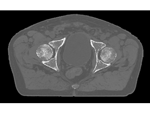
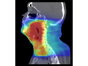
As a plug-in clinical application to Ingenia MR-RT, MRCAT Prostate + Auto-Contouring provides attenuation maps and automated, MR-based contours of prostate and organs at risk in as little as 20 minutes – all in a repeatable ‘one-click’ workflow.

Features
Drive speed, accuracy and consistency

Drive speed, accuracy and consistency

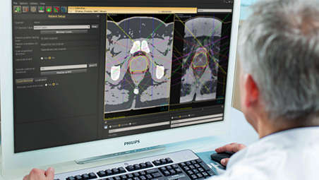
Because MRCAT Prostate requires input from MR images only, it reduces the organization and coordination of scans, eliminates the effort involved in MR-CT registration, and saves the patient from undergoing multiple procedures. Moreover, Auto-Contouring automates standard, labor-intensive and repetitive tasks, while at the same time reducing variability and errors caused by manual steps. This improves consistency and reproducibility - for more confidence in the planning process.

Drive speed, accuracy and consistency
Drive speed, accuracy and consistency
Because MRCAT Prostate requires input from MR images only, it reduces the organization and coordination of scans, eliminates the effort involved in MR-CT registration, and saves the patient from undergoing multiple procedures. Moreover, Auto-Contouring automates standard, labor-intensive and repetitive tasks, while at the same time reducing variability and errors caused by manual steps. This improves consistency and reproducibility - for more confidence in the planning process.

Drive speed, accuracy and consistency

Because MRCAT Prostate requires input from MR images only, it reduces the organization and coordination of scans, eliminates the effort involved in MR-CT registration, and saves the patient from undergoing multiple procedures. Moreover, Auto-Contouring automates standard, labor-intensive and repetitive tasks, while at the same time reducing variability and errors caused by manual steps. This improves consistency and reproducibility - for more confidence in the planning process.
Click here for more information
Drive speed, accuracy and consistency

Drive speed, accuracy and consistency

Because MRCAT Prostate requires input from MR images only, it reduces the organization and coordination of scans, eliminates the effort involved in MR-CT registration, and saves the patient from undergoing multiple procedures. Moreover, Auto-Contouring automates standard, labor-intensive and repetitive tasks, while at the same time reducing variability and errors caused by manual steps. This improves consistency and reproducibility - for more confidence in the planning process.
Fast, consistent imaging protocol
Fast, consistent imaging protocol

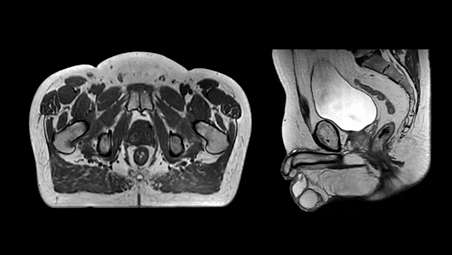
A dedicated, standardized imaging protocol includes a T1W mDIXON XD and a T2W TSE scan as source data for the generation of MRCAT (MR for Calculating ATtenuation) density maps and MR-based Auto-Contouring. Compressed SENSE acceleration keeps the total scan time short, which promotes patient comfort by minimizing time in the scanner and helps to boost productivity.

Fast, consistent imaging protocol

Fast, consistent imaging protocol
A dedicated, standardized imaging protocol includes a T1W mDIXON XD and a T2W TSE scan as source data for the generation of MRCAT (MR for Calculating ATtenuation) density maps and MR-based Auto-Contouring. Compressed SENSE acceleration keeps the total scan time short, which promotes patient comfort by minimizing time in the scanner and helps to boost productivity.

Fast, consistent imaging protocol

A dedicated, standardized imaging protocol includes a T1W mDIXON XD and a T2W TSE scan as source data for the generation of MRCAT (MR for Calculating ATtenuation) density maps and MR-based Auto-Contouring. Compressed SENSE acceleration keeps the total scan time short, which promotes patient comfort by minimizing time in the scanner and helps to boost productivity.
Click here for more information
Fast, consistent imaging protocol

Fast, consistent imaging protocol

A dedicated, standardized imaging protocol includes a T1W mDIXON XD and a T2W TSE scan as source data for the generation of MRCAT (MR for Calculating ATtenuation) density maps and MR-based Auto-Contouring. Compressed SENSE acceleration keeps the total scan time short, which promotes patient comfort by minimizing time in the scanner and helps to boost productivity.
Automatic generation of synthetic CT images

Automatic generation of synthetic CT images

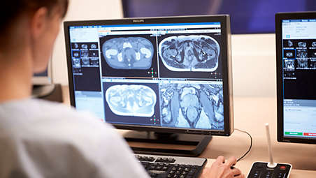
MRCAT Prostate automatically generates attenuation maps using the high-resolution mDIXON scan as source. Smart, validated algorithms enable automatic tissue segmentation and assignment of Hounsfield Units to deliver MRCAT images with CT-like density information for dose calculations - directly on the MR console.

Automatic generation of synthetic CT images

Automatic generation of synthetic CT images
MRCAT Prostate automatically generates attenuation maps using the high-resolution mDIXON scan as source. Smart, validated algorithms enable automatic tissue segmentation and assignment of Hounsfield Units to deliver MRCAT images with CT-like density information for dose calculations - directly on the MR console.

Automatic generation of synthetic CT images

MRCAT Prostate automatically generates attenuation maps using the high-resolution mDIXON scan as source. Smart, validated algorithms enable automatic tissue segmentation and assignment of Hounsfield Units to deliver MRCAT images with CT-like density information for dose calculations - directly on the MR console.
Click here for more information
Automatic generation of synthetic CT images

Automatic generation of synthetic CT images

MRCAT Prostate automatically generates attenuation maps using the high-resolution mDIXON scan as source. Smart, validated algorithms enable automatic tissue segmentation and assignment of Hounsfield Units to deliver MRCAT images with CT-like density information for dose calculations - directly on the MR console.
Accuracy in dose planning
Accuracy in dose planning

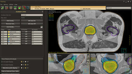
The MRCAT Prostate scanning protocol and generation algorithms have been designed with the strict accuracy requirements of RT in mind. MRCAT Prostate images have high geometric accuracy* and validation studies have shown that MRCAT-based dose plans are robust and equivalent** to CT-based plans promoting confidence in dose planning.

Accuracy in dose planning

Accuracy in dose planning
The MRCAT Prostate scanning protocol and generation algorithms have been designed with the strict accuracy requirements of RT in mind. MRCAT Prostate images have high geometric accuracy* and validation studies have shown that MRCAT-based dose plans are robust and equivalent** to CT-based plans promoting confidence in dose planning.

Accuracy in dose planning

The MRCAT Prostate scanning protocol and generation algorithms have been designed with the strict accuracy requirements of RT in mind. MRCAT Prostate images have high geometric accuracy* and validation studies have shown that MRCAT-based dose plans are robust and equivalent** to CT-based plans promoting confidence in dose planning.
Click here for more information
Accuracy in dose planning

Accuracy in dose planning

The MRCAT Prostate scanning protocol and generation algorithms have been designed with the strict accuracy requirements of RT in mind. MRCAT Prostate images have high geometric accuracy* and validation studies have shown that MRCAT-based dose plans are robust and equivalent** to CT-based plans promoting confidence in dose planning.
Create accurate*** contours with little to no user interaction

Create accurate*** contours with little to no user interaction

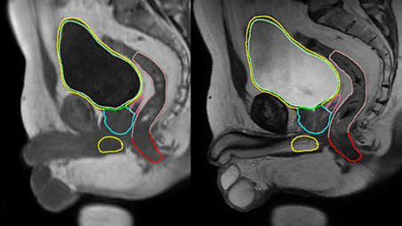
MR-based Auto-Contouring automatically creates contours of prostate and OARs, reducing repetitive tasks and time spent, compared to manual methods. It uses dedicated MR imaging data based on the 3D T2W TSE and T1W mDIXON XD sequences and model-based adaptive algorithms. Auto-Contouring delineation of prostate and OARs has been found to be accurate (average distance < 1.5mm)*** in at least 70% of contours evaluated****. This significantly reduces the need for manual contouring or manual adaptations, while increasing consistency.

Create accurate*** contours with little to no user interaction

Create accurate*** contours with little to no user interaction
MR-based Auto-Contouring automatically creates contours of prostate and OARs, reducing repetitive tasks and time spent, compared to manual methods. It uses dedicated MR imaging data based on the 3D T2W TSE and T1W mDIXON XD sequences and model-based adaptive algorithms. Auto-Contouring delineation of prostate and OARs has been found to be accurate (average distance < 1.5mm)*** in at least 70% of contours evaluated****. This significantly reduces the need for manual contouring or manual adaptations, while increasing consistency.

Create accurate*** contours with little to no user interaction

MR-based Auto-Contouring automatically creates contours of prostate and OARs, reducing repetitive tasks and time spent, compared to manual methods. It uses dedicated MR imaging data based on the 3D T2W TSE and T1W mDIXON XD sequences and model-based adaptive algorithms. Auto-Contouring delineation of prostate and OARs has been found to be accurate (average distance < 1.5mm)*** in at least 70% of contours evaluated****. This significantly reduces the need for manual contouring or manual adaptations, while increasing consistency.
Click here for more information
Create accurate*** contours with little to no user interaction

Create accurate*** contours with little to no user interaction

MR-based Auto-Contouring automatically creates contours of prostate and OARs, reducing repetitive tasks and time spent, compared to manual methods. It uses dedicated MR imaging data based on the 3D T2W TSE and T1W mDIXON XD sequences and model-based adaptive algorithms. Auto-Contouring delineation of prostate and OARs has been found to be accurate (average distance < 1.5mm)*** in at least 70% of contours evaluated****. This significantly reduces the need for manual contouring or manual adaptations, while increasing consistency.
MRI as primary image set in treatment planning

MRI as primary image set in treatment planning

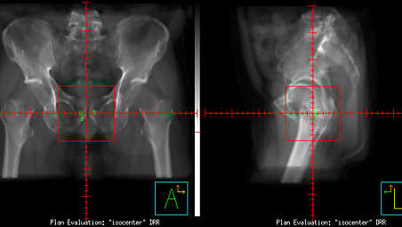
The MRCAT images generated on the MR console conform to the DICOM standard (modality CT) and hence can be exported to treatment planning systems (TPS) as the primary image dataset. Together with the generated contours (RTSTRUCT) and the ability to generate MRCAT-based digitally reconstructed radiographs (DRRs), you can replace your traditional CT-based workflow with an MRI-only radiotherapy workflow from imaging and planning to position verification.

MRI as primary image set in treatment planning

MRI as primary image set in treatment planning
The MRCAT images generated on the MR console conform to the DICOM standard (modality CT) and hence can be exported to treatment planning systems (TPS) as the primary image dataset. Together with the generated contours (RTSTRUCT) and the ability to generate MRCAT-based digitally reconstructed radiographs (DRRs), you can replace your traditional CT-based workflow with an MRI-only radiotherapy workflow from imaging and planning to position verification.

MRI as primary image set in treatment planning

The MRCAT images generated on the MR console conform to the DICOM standard (modality CT) and hence can be exported to treatment planning systems (TPS) as the primary image dataset. Together with the generated contours (RTSTRUCT) and the ability to generate MRCAT-based digitally reconstructed radiographs (DRRs), you can replace your traditional CT-based workflow with an MRI-only radiotherapy workflow from imaging and planning to position verification.
Click here for more information
MRI as primary image set in treatment planning

MRI as primary image set in treatment planning

The MRCAT images generated on the MR console conform to the DICOM standard (modality CT) and hence can be exported to treatment planning systems (TPS) as the primary image dataset. Together with the generated contours (RTSTRUCT) and the ability to generate MRCAT-based digitally reconstructed radiographs (DRRs), you can replace your traditional CT-based workflow with an MRI-only radiotherapy workflow from imaging and planning to position verification.
Put Philips MR-only radiotherapy to work today

Put Philips MR-only radiotherapy to work today

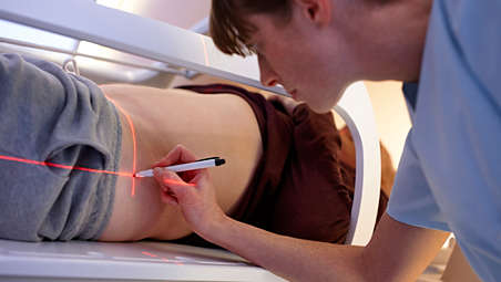
To successfully bring MR-only radiotherapy into your clinical routine, we recognize that you must look beyond the imaging itself and address important steps such as patient marking, position verification, and quality assurance. We are prepared to support you throughout this process. To this end, we offer dedicated workflow descriptions, best practice sharing and tailored training support, designed to provide assistance as you adopt this new treatment paradigm.

Put Philips MR-only radiotherapy to work today

Put Philips MR-only radiotherapy to work today
To successfully bring MR-only radiotherapy into your clinical routine, we recognize that you must look beyond the imaging itself and address important steps such as patient marking, position verification, and quality assurance. We are prepared to support you throughout this process. To this end, we offer dedicated workflow descriptions, best practice sharing and tailored training support, designed to provide assistance as you adopt this new treatment paradigm.

Put Philips MR-only radiotherapy to work today

To successfully bring MR-only radiotherapy into your clinical routine, we recognize that you must look beyond the imaging itself and address important steps such as patient marking, position verification, and quality assurance. We are prepared to support you throughout this process. To this end, we offer dedicated workflow descriptions, best practice sharing and tailored training support, designed to provide assistance as you adopt this new treatment paradigm.
Click here for more information
Put Philips MR-only radiotherapy to work today

Put Philips MR-only radiotherapy to work today

To successfully bring MR-only radiotherapy into your clinical routine, we recognize that you must look beyond the imaging itself and address important steps such as patient marking, position verification, and quality assurance. We are prepared to support you throughout this process. To this end, we offer dedicated workflow descriptions, best practice sharing and tailored training support, designed to provide assistance as you adopt this new treatment paradigm.
  • Drive speed, accuracy and consistency
  • Fast, consistent imaging protocol
  • Automatic generation of synthetic CT images
  • Accuracy in dose planning
See all features
Drive speed, accuracy and consistency

Drive speed, accuracy and consistency

Because MRCAT Prostate requires input from MR images only, it reduces the organization and coordination of scans, eliminates the effort involved in MR-CT registration, and saves the patient from undergoing multiple procedures. Moreover, Auto-Contouring automates standard, labor-intensive and repetitive tasks, while at the same time reducing variability and errors caused by manual steps. This improves consistency and reproducibility - for more confidence in the planning process.

Drive speed, accuracy and consistency

Drive speed, accuracy and consistency
Because MRCAT Prostate requires input from MR images only, it reduces the organization and coordination of scans, eliminates the effort involved in MR-CT registration, and saves the patient from undergoing multiple procedures. Moreover, Auto-Contouring automates standard, labor-intensive and repetitive tasks, while at the same time reducing variability and errors caused by manual steps. This improves consistency and reproducibility - for more confidence in the planning process.

Drive speed, accuracy and consistency

Because MRCAT Prostate requires input from MR images only, it reduces the organization and coordination of scans, eliminates the effort involved in MR-CT registration, and saves the patient from undergoing multiple procedures. Moreover, Auto-Contouring automates standard, labor-intensive and repetitive tasks, while at the same time reducing variability and errors caused by manual steps. This improves consistency and reproducibility - for more confidence in the planning process.
Click here for more information
Drive speed, accuracy and consistency

Drive speed, accuracy and consistency

Because MRCAT Prostate requires input from MR images only, it reduces the organization and coordination of scans, eliminates the effort involved in MR-CT registration, and saves the patient from undergoing multiple procedures. Moreover, Auto-Contouring automates standard, labor-intensive and repetitive tasks, while at the same time reducing variability and errors caused by manual steps. This improves consistency and reproducibility - for more confidence in the planning process.
Fast, consistent imaging protocol

Fast, consistent imaging protocol

A dedicated, standardized imaging protocol includes a T1W mDIXON XD and a T2W TSE scan as source data for the generation of MRCAT (MR for Calculating ATtenuation) density maps and MR-based Auto-Contouring. Compressed SENSE acceleration keeps the total scan time short, which promotes patient comfort by minimizing time in the scanner and helps to boost productivity.

Fast, consistent imaging protocol

Fast, consistent imaging protocol
A dedicated, standardized imaging protocol includes a T1W mDIXON XD and a T2W TSE scan as source data for the generation of MRCAT (MR for Calculating ATtenuation) density maps and MR-based Auto-Contouring. Compressed SENSE acceleration keeps the total scan time short, which promotes patient comfort by minimizing time in the scanner and helps to boost productivity.

Fast, consistent imaging protocol

A dedicated, standardized imaging protocol includes a T1W mDIXON XD and a T2W TSE scan as source data for the generation of MRCAT (MR for Calculating ATtenuation) density maps and MR-based Auto-Contouring. Compressed SENSE acceleration keeps the total scan time short, which promotes patient comfort by minimizing time in the scanner and helps to boost productivity.
Click here for more information
Fast, consistent imaging protocol

Fast, consistent imaging protocol

A dedicated, standardized imaging protocol includes a T1W mDIXON XD and a T2W TSE scan as source data for the generation of MRCAT (MR for Calculating ATtenuation) density maps and MR-based Auto-Contouring. Compressed SENSE acceleration keeps the total scan time short, which promotes patient comfort by minimizing time in the scanner and helps to boost productivity.
Automatic generation of synthetic CT images

Automatic generation of synthetic CT images

MRCAT Prostate automatically generates attenuation maps using the high-resolution mDIXON scan as source. Smart, validated algorithms enable automatic tissue segmentation and assignment of Hounsfield Units to deliver MRCAT images with CT-like density information for dose calculations - directly on the MR console.

Automatic generation of synthetic CT images

Automatic generation of synthetic CT images
MRCAT Prostate automatically generates attenuation maps using the high-resolution mDIXON scan as source. Smart, validated algorithms enable automatic tissue segmentation and assignment of Hounsfield Units to deliver MRCAT images with CT-like density information for dose calculations - directly on the MR console.

Automatic generation of synthetic CT images

MRCAT Prostate automatically generates attenuation maps using the high-resolution mDIXON scan as source. Smart, validated algorithms enable automatic tissue segmentation and assignment of Hounsfield Units to deliver MRCAT images with CT-like density information for dose calculations - directly on the MR console.
Click here for more information
Automatic generation of synthetic CT images

Automatic generation of synthetic CT images

MRCAT Prostate automatically generates attenuation maps using the high-resolution mDIXON scan as source. Smart, validated algorithms enable automatic tissue segmentation and assignment of Hounsfield Units to deliver MRCAT images with CT-like density information for dose calculations - directly on the MR console.
Accuracy in dose planning

Accuracy in dose planning

The MRCAT Prostate scanning protocol and generation algorithms have been designed with the strict accuracy requirements of RT in mind. MRCAT Prostate images have high geometric accuracy* and validation studies have shown that MRCAT-based dose plans are robust and equivalent** to CT-based plans promoting confidence in dose planning.

Accuracy in dose planning

Accuracy in dose planning
The MRCAT Prostate scanning protocol and generation algorithms have been designed with the strict accuracy requirements of RT in mind. MRCAT Prostate images have high geometric accuracy* and validation studies have shown that MRCAT-based dose plans are robust and equivalent** to CT-based plans promoting confidence in dose planning.

Accuracy in dose planning

The MRCAT Prostate scanning protocol and generation algorithms have been designed with the strict accuracy requirements of RT in mind. MRCAT Prostate images have high geometric accuracy* and validation studies have shown that MRCAT-based dose plans are robust and equivalent** to CT-based plans promoting confidence in dose planning.
Click here for more information
Accuracy in dose planning

Accuracy in dose planning

The MRCAT Prostate scanning protocol and generation algorithms have been designed with the strict accuracy requirements of RT in mind. MRCAT Prostate images have high geometric accuracy* and validation studies have shown that MRCAT-based dose plans are robust and equivalent** to CT-based plans promoting confidence in dose planning.
Create accurate*** contours with little to no user interaction

Create accurate*** contours with little to no user interaction

MR-based Auto-Contouring automatically creates contours of prostate and OARs, reducing repetitive tasks and time spent, compared to manual methods. It uses dedicated MR imaging data based on the 3D T2W TSE and T1W mDIXON XD sequences and model-based adaptive algorithms. Auto-Contouring delineation of prostate and OARs has been found to be accurate (average distance < 1.5mm)*** in at least 70% of contours evaluated****. This significantly reduces the need for manual contouring or manual adaptations, while increasing consistency.

Create accurate*** contours with little to no user interaction

Create accurate*** contours with little to no user interaction
MR-based Auto-Contouring automatically creates contours of prostate and OARs, reducing repetitive tasks and time spent, compared to manual methods. It uses dedicated MR imaging data based on the 3D T2W TSE and T1W mDIXON XD sequences and model-based adaptive algorithms. Auto-Contouring delineation of prostate and OARs has been found to be accurate (average distance < 1.5mm)*** in at least 70% of contours evaluated****. This significantly reduces the need for manual contouring or manual adaptations, while increasing consistency.

Create accurate*** contours with little to no user interaction

MR-based Auto-Contouring automatically creates contours of prostate and OARs, reducing repetitive tasks and time spent, compared to manual methods. It uses dedicated MR imaging data based on the 3D T2W TSE and T1W mDIXON XD sequences and model-based adaptive algorithms. Auto-Contouring delineation of prostate and OARs has been found to be accurate (average distance < 1.5mm)*** in at least 70% of contours evaluated****. This significantly reduces the need for manual contouring or manual adaptations, while increasing consistency.
Click here for more information
Create accurate*** contours with little to no user interaction

Create accurate*** contours with little to no user interaction

MR-based Auto-Contouring automatically creates contours of prostate and OARs, reducing repetitive tasks and time spent, compared to manual methods. It uses dedicated MR imaging data based on the 3D T2W TSE and T1W mDIXON XD sequences and model-based adaptive algorithms. Auto-Contouring delineation of prostate and OARs has been found to be accurate (average distance < 1.5mm)*** in at least 70% of contours evaluated****. This significantly reduces the need for manual contouring or manual adaptations, while increasing consistency.
MRI as primary image set in treatment planning

MRI as primary image set in treatment planning

The MRCAT images generated on the MR console conform to the DICOM standard (modality CT) and hence can be exported to treatment planning systems (TPS) as the primary image dataset. Together with the generated contours (RTSTRUCT) and the ability to generate MRCAT-based digitally reconstructed radiographs (DRRs), you can replace your traditional CT-based workflow with an MRI-only radiotherapy workflow from imaging and planning to position verification.

MRI as primary image set in treatment planning

MRI as primary image set in treatment planning
The MRCAT images generated on the MR console conform to the DICOM standard (modality CT) and hence can be exported to treatment planning systems (TPS) as the primary image dataset. Together with the generated contours (RTSTRUCT) and the ability to generate MRCAT-based digitally reconstructed radiographs (DRRs), you can replace your traditional CT-based workflow with an MRI-only radiotherapy workflow from imaging and planning to position verification.

MRI as primary image set in treatment planning

The MRCAT images generated on the MR console conform to the DICOM standard (modality CT) and hence can be exported to treatment planning systems (TPS) as the primary image dataset. Together with the generated contours (RTSTRUCT) and the ability to generate MRCAT-based digitally reconstructed radiographs (DRRs), you can replace your traditional CT-based workflow with an MRI-only radiotherapy workflow from imaging and planning to position verification.
Click here for more information
MRI as primary image set in treatment planning

MRI as primary image set in treatment planning

The MRCAT images generated on the MR console conform to the DICOM standard (modality CT) and hence can be exported to treatment planning systems (TPS) as the primary image dataset. Together with the generated contours (RTSTRUCT) and the ability to generate MRCAT-based digitally reconstructed radiographs (DRRs), you can replace your traditional CT-based workflow with an MRI-only radiotherapy workflow from imaging and planning to position verification.
Put Philips MR-only radiotherapy to work today

Put Philips MR-only radiotherapy to work today

To successfully bring MR-only radiotherapy into your clinical routine, we recognize that you must look beyond the imaging itself and address important steps such as patient marking, position verification, and quality assurance. We are prepared to support you throughout this process. To this end, we offer dedicated workflow descriptions, best practice sharing and tailored training support, designed to provide assistance as you adopt this new treatment paradigm.

Put Philips MR-only radiotherapy to work today

Put Philips MR-only radiotherapy to work today
To successfully bring MR-only radiotherapy into your clinical routine, we recognize that you must look beyond the imaging itself and address important steps such as patient marking, position verification, and quality assurance. We are prepared to support you throughout this process. To this end, we offer dedicated workflow descriptions, best practice sharing and tailored training support, designed to provide assistance as you adopt this new treatment paradigm.

Put Philips MR-only radiotherapy to work today

To successfully bring MR-only radiotherapy into your clinical routine, we recognize that you must look beyond the imaging itself and address important steps such as patient marking, position verification, and quality assurance. We are prepared to support you throughout this process. To this end, we offer dedicated workflow descriptions, best practice sharing and tailored training support, designed to provide assistance as you adopt this new treatment paradigm.
Click here for more information
Put Philips MR-only radiotherapy to work today

Put Philips MR-only radiotherapy to work today

To successfully bring MR-only radiotherapy into your clinical routine, we recognize that you must look beyond the imaging itself and address important steps such as patient marking, position verification, and quality assurance. We are prepared to support you throughout this process. To this end, we offer dedicated workflow descriptions, best practice sharing and tailored training support, designed to provide assistance as you adopt this new treatment paradigm.

Specifications

MRCAT Prostate and Auto-Contouring
Compatibility MR system
  • Ingenia 1.5T and 3.0T MR-RT, Ambition 1.5T MR-RT and Elition 3.0T MR-RT
MRCAT Prostate and Auto-Contouring
Compatibility MR system
  • Ingenia 1.5T and 3.0T MR-RT, Ambition 1.5T MR-RT and Elition 3.0T MR-RT
See all specifications
MRCAT Prostate and Auto-Contouring
Compatibility MR system
  • Ingenia 1.5T and 3.0T MR-RT, Ambition 1.5T MR-RT and Elition 3.0T MR-RT
  • *Accurate means: MRCAT provides < ± 1 mm total geometric accuracy of image data in < 20 cm Diameter Spherical Volume (DSV) and < ± 2 mm total geometric accuracy of image data in < 40 cm Diameter Spherical Volume (DSV))*. *Limited to 32 cm in z-direction in more than 95% of the points within the volume
  • **The simulated dose based on MRCAT images does not differ in >95% of prostate cancer patients (Gamma analysis criterion 3%/3 mm realized in 99% of voxels exceeding 75% of the maximum dose) when compared with the CT-based plan for EBRT.
  • ***Accurate means 95th percentile modified Hausdorff distance <5mm compared to contours made by experts manually. Average distance is <1.5 mm and is measured as average modified Hausdorff distance compared to contours made by experts manually.
  • **** Based on 49 cases (each for anatomical prostate, bladder, rectum, penile bulb and femur heads)
You are about to visit a Philips global content page

Continue

You are about to visit a Philips global content page

Continue

Our site can best be viewed with the latest version of Microsoft Edge, Google Chrome or Firefox.

Jūs ieejat Latvijas Philips Healthcare tīmekļa vietnē.
Please select the checkbox

Tīmekļa vietne ir paredzēta tikai personām, kuras izmanto medicīniskās ierīces kā profesionāļi, tostarp personām, kas praktizē medicīnas profesijās, personām, kas darbojas veselības aprūpes iestāžu labā vai organizācijām, kas tirgo medicīnas ierīces kā profesionāļi.

Vai esat persona, kas atbilst iepriekš minētajiem kritērijiem?