- Unleash the real power of MR simulation
-
Unleash the real power of MR simulation
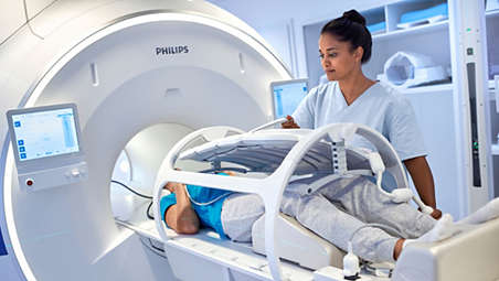
MRCAT Pelvis lets you plan radiation therapy for male and female pelvic cancer patients with soft-tissue tumors using MRI as a single-modality solution. This not only extends the benefits of MRI’s outstanding soft-tissue contrast to radiotherapy planning, but it also eliminates arduous, error-prone CT-MRI registration from the process, reducing uncertainties and complexity. - Fast, consistent imaging protocol
-
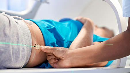
Fast, consistent imaging protocol
The dedicated MRCAT Pelvis imaging protocol includes a single, high-resolution, multi-contrast mDIXON sequence as the source for MRCAT generation. This scan is accelerated by Compressed SENSE, promoting patient comfort by minimizing time in the scanner. Moreover, it is standardized to deliver consistent results. A complementary 3D T2W scan provides high geometric accuracy and high-resolution image quality to support accurate delineation of target and critical structures. The total imaging protocol takes less than 15 minutes. - Automatic generation of synthetic CT images
-
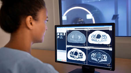
Automatic generation of synthetic CT images
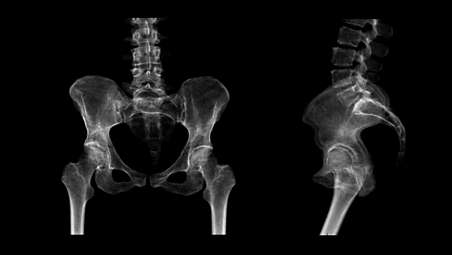
MRCAT images are automatically generated using the mDIXON scan as source. Embedded image post-processing runs in the background, parallel to image acquisition, adding no time to the scanning session. Smart, validated algorithms enable automatic tissue segmentation and assignment of continuous Hounsfield units to deliver MRCAT images with CT-like density information for dose calculations. - Accuracy in dose planning
-
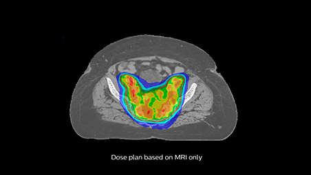
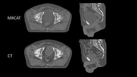
Accuracy in dose planning
MRCAT images have high geometric accuracy* and validation studies have shown that MRCAT-based dose plans are robust and as accurate** as CT-based plans promoting confidence in dose planning. - Patient positioning based on MR-only imaging
-
Patient positioning based on MR-only imaging
The MR-based image sets with continuous Hounsfield units enable CBCT-based positioning based on soft-tissue contrast with the look and feel of CT. You can also use MRCAT data to generate MR-based digitally reconstructed radiographs (DRRs) to allow for patient positioning using bony structure. - Put Philips MR-only radiotherapy to work today
-
Put Philips MR-only radiotherapy to work today
To successfully bring MR-only radiotherapy into your clinical routine, we recognize that you must look beyond the imaging itself and address important steps such as patient marking, position verification, and quality assurance. We are prepared to support you throughout this process. To this end, we offer dedicated workflow descriptions, best practice sharing and tailored training support, designed to provide assistance as you adopt this new treatment paradigm.
Unleash the real power of MR simulation
Unleash the real power of MR simulation
Unleash the real power of MR simulation
Fast, consistent imaging protocol
Fast, consistent imaging protocol
Fast, consistent imaging protocol
Automatic generation of synthetic CT images
Automatic generation of synthetic CT images
Automatic generation of synthetic CT images
Accuracy in dose planning
Accuracy in dose planning
Accuracy in dose planning
Patient positioning based on MR-only imaging
Patient positioning based on MR-only imaging
Patient positioning based on MR-only imaging
Put Philips MR-only radiotherapy to work today
Put Philips MR-only radiotherapy to work today
Put Philips MR-only radiotherapy to work today
- Unleash the real power of MR simulation
- Fast, consistent imaging protocol
- Automatic generation of synthetic CT images
- Accuracy in dose planning
- Unleash the real power of MR simulation
-
Unleash the real power of MR simulation
MRCAT Pelvis lets you plan radiation therapy for male and female pelvic cancer patients with soft-tissue tumors using MRI as a single-modality solution. This not only extends the benefits of MRI’s outstanding soft-tissue contrast to radiotherapy planning, but it also eliminates arduous, error-prone CT-MRI registration from the process, reducing uncertainties and complexity. - Fast, consistent imaging protocol
-
Fast, consistent imaging protocol
The dedicated MRCAT Pelvis imaging protocol includes a single, high-resolution, multi-contrast mDIXON sequence as the source for MRCAT generation. This scan is accelerated by Compressed SENSE, promoting patient comfort by minimizing time in the scanner. Moreover, it is standardized to deliver consistent results. A complementary 3D T2W scan provides high geometric accuracy and high-resolution image quality to support accurate delineation of target and critical structures. The total imaging protocol takes less than 15 minutes. - Automatic generation of synthetic CT images
-
Automatic generation of synthetic CT images
MRCAT images are automatically generated using the mDIXON scan as source. Embedded image post-processing runs in the background, parallel to image acquisition, adding no time to the scanning session. Smart, validated algorithms enable automatic tissue segmentation and assignment of continuous Hounsfield units to deliver MRCAT images with CT-like density information for dose calculations. - Accuracy in dose planning
-
Accuracy in dose planning
MRCAT images have high geometric accuracy* and validation studies have shown that MRCAT-based dose plans are robust and as accurate** as CT-based plans promoting confidence in dose planning. - Patient positioning based on MR-only imaging
-
Patient positioning based on MR-only imaging
The MR-based image sets with continuous Hounsfield units enable CBCT-based positioning based on soft-tissue contrast with the look and feel of CT. You can also use MRCAT data to generate MR-based digitally reconstructed radiographs (DRRs) to allow for patient positioning using bony structure. - Put Philips MR-only radiotherapy to work today
-
Put Philips MR-only radiotherapy to work today
To successfully bring MR-only radiotherapy into your clinical routine, we recognize that you must look beyond the imaging itself and address important steps such as patient marking, position verification, and quality assurance. We are prepared to support you throughout this process. To this end, we offer dedicated workflow descriptions, best practice sharing and tailored training support, designed to provide assistance as you adopt this new treatment paradigm.
Unleash the real power of MR simulation
Unleash the real power of MR simulation
Unleash the real power of MR simulation
Fast, consistent imaging protocol
Fast, consistent imaging protocol
Fast, consistent imaging protocol
Automatic generation of synthetic CT images
Automatic generation of synthetic CT images
Automatic generation of synthetic CT images
Accuracy in dose planning
Accuracy in dose planning
Accuracy in dose planning
Patient positioning based on MR-only imaging
Patient positioning based on MR-only imaging
Patient positioning based on MR-only imaging
Put Philips MR-only radiotherapy to work today
Put Philips MR-only radiotherapy to work today
Put Philips MR-only radiotherapy to work today
Specifications
- MRCAT Pelvis
-
MRCAT Pelvis Compatibility MR system - Ingenia 1.5T and 3.0T MR-RT, Ambition 1.5T MR-RT and Elition 3.0T MR-RT
-
- MRCAT Pelvis
-
MRCAT Pelvis Compatibility MR system - Ingenia 1.5T and 3.0T MR-RT, Ambition 1.5T MR-RT and Elition 3.0T MR-RT
-
- MRCAT Pelvis
-
MRCAT Pelvis Compatibility MR system - Ingenia 1.5T and 3.0T MR-RT, Ambition 1.5T MR-RT and Elition 3.0T MR-RT
-
Related products
Alternative products
-
Ingenia MR-RT XD
- Experience the MRI difference
- Position with precision
- Coil solutions for RT imaging
- MR-only radiotherapy planning
View product
-
MRCAT Brain
- MR-only sim for primary and metastatic tumors in the brain
- Single-scan approach
- Automatic generation of synthetic CT images using AI
- Accuracy in dose planning
View product
-
MRCAT Prostate + Auto-Contouring
- MR-sim and contouring in 20 minutes
- Automatic generation of synthetic CT images
- Accurate contours with little to no user interaction
- Accuracy in dose planning
View product
-
MRCAT Head and Neck
- MR-only sim for soft tissue tumors in the head and neck region
- Short scan times promote patient comfort
- Automatic generation of CT-like density information using AI
- Accuracy in dose planning
View product


-
Ingenia MR-RT XD
The Philips Ingenia MR-RT XD platform harnesses the power and value of MRI for radiation therapy planning. It has been designed around the needs of radiation oncology, with ease-of-use, streamlined integration, and versatility in mind. Central to that concept is the ability to define a tailored approach with customizable functionality that meets your individual clinical, workflow, and budgetary requirements – all to provide better patient care.
View product
-
MRCAT Brain
MRCAT Brain clinical application allows the use of MRI as the primary imaging modality for radiotherapy planning of primary and metastatic tumors in the brain without the need for CT. Detailed anatomical information for contouring and attenuation maps for dose calculations are both obtained from a single, submillimeter resolution 3D T1W mDIXON MR sequence. Artificial Intelligence (AI) is used for fast computation of continuous Hounsfield units directly on the MR console.
View product
-
MRCAT Prostate + Auto-Contouring
As a plug-in clinical application to Ingenia MR-RT, MRCAT Prostate + Auto-Contouring provides attenuation maps and automated, MR-based contours of prostate and organs at risk in as little as 20 minutes – all in a repeatable ‘one-click’ workflow.
View product
See all related products
- *Accurate means: MRCAT image acquisition provides < ± 1 mm geometric accuracy of image data in < 20 cm Diameter Spherical Volume (DSV) and < ± 2 mm geometric accuracy of image data in < 40 cm Diameter Spherical Volume (DSV)*. * Limited to 32 cm in z-direction in more than 95% of the points within the volume
- **The simulated dose based on MRCAT images does not differ (Gamma analysis criterion 3%/3mm realized in 99% of voxels within the PTV or exceeding 75% of the maximum dose) in 95% of the pelvic cancer patients when compared with CT-based plan for EBRT.