- Drive the precision of radiation therapy
-
Drive the precision of radiation therapy
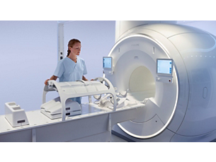
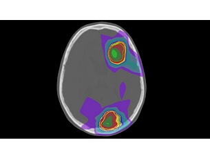
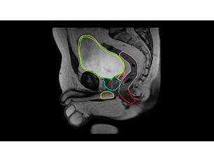
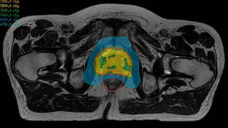
Whether for external beam radiation therapy (RT) or brachytherapy, integrating MR imaging into CT‑based planning can harness the power of MRI and transform patient management. With MRI’s excellent soft-tissue contrast, you can clearly see the tumor and organs at risk. So you can support accuracy in delineation and design the best possible treatment plans. Image courtesy of William Beaumont Health System, Detroit, USA - A superb MRI platform for radiation oncology
-
A superb MRI platform for radiation oncology
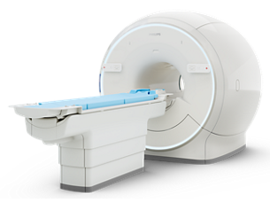
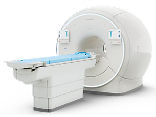
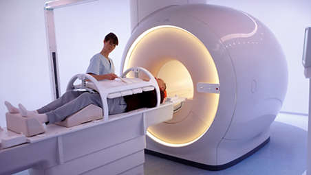
Ingenia MR-RT drives clinical excellence with state-of-the art image quality and high geometric accuracy thanks to dStream architecture, high gradient linearity, and 3D Gradient Distortion Correction. With the state-of-the art next generation Elition 3.0T and Ambition 1.5T wide-bore MR systems, you can benefit from MRI innovations, now and in years to come. - Maintain high standards
-
Maintain high standards
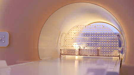
Know you can rely on MRI performance. Evaluate the geometric accuracy in a large field of view with the ready-to-use QA package that includes a phantom and analysis software. Most steps are fully automated, so you can perform routine volumetric evaluations fast and in a repeatable manner. The on-console Pass/Fail analysis provides users with clear guidance on the outcome of the geometric accuracy analysis. The result is user independent and unambiguous. - Position with precision
-
Position with precision
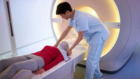
Highly-targeted RT plans rely on reproducible patient positioning in the treatment position. Unique to Philips, the integrated MR-RT CouchTop frees up in-bore space while improving SNR by bringing patients closer to the posterior coil*. Complete with indexing, the CouchTop accommodates a variety of MRI-compatible immobilization accessories from main vendors. - Work your way
-
Work your way
Refine workflows with a system that fits how you work. The optional LAP DORADOnova MR3T laser positioning system supports enhanced MR-CT registration since it allows you to align patients at the MRI scanner. One-click travel-to-scan moves patients directly to the MRI system isocenter after laser alignment, thereby reducing workflow steps - Set up easily and flexibly
-
Set up easily and flexibly
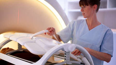
The Anterior Coil Support enables easy and flexible coil setup with large bore access and space for patient immobilization. The support can be easily tilted by a single operator to bring the coil close to the patient to optimize SNR without touching the body’s contours. - MR-linac simulation package for Elekta Unity
-
MR-linac simulation package for Elekta Unity
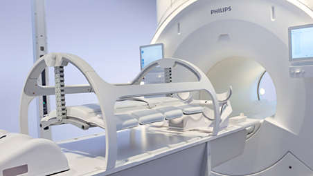
The Philips Ingenia MR-RT simulation platform with MR-linac simulation package is an ideal complement to Elekta Unity. With consistent workflows and image quality from MR simulation through to online MR guidance during radiation treatment, it will let you exploit the many similarities and synergies between Philips Ingenia MR-RT and Elekta Unity. - See clearly in treatment planning
-
See clearly in treatment planning
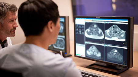
Enjoy consistent, excellent image quality for multiple anatomies. Versatile arrangements of dStream coils work together with ExamCards tailored for RT to provide high-contrast images with high geometric fidelity. Quickly execute complete imaging protocols for prostate, female pelvis, brain, head and neck, and spine. - Learn and share MRI expertise
-
Learn and share MRI expertise
Successful integration of MR imaging in your workflow starts with people. We offer tailored training to assist your team in streamlining workflows and making full, efficient use of MR imaging from day one. - MR-only radiotherapy
-
MR-only radiotherapy
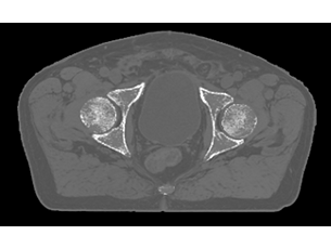
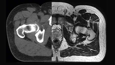
Our innovative MRCAT (MR for Calculating ATtenuation) clinical applications lets you plan radiation therapy using MRI as primary imaging modality. Within just one, fast MR exam, MRCAT provides both excellent soft-tissue contrast for target and OAR delineation and CT-like density information for dose calculations. This not only extends the benefits of MRI’s excellent soft-tissue contrast to radiotherapy planning, but it also eliminates arduous, error-prone CT-MRI registration from the process, reducing uncertainties and complexity. Check out the related product section for the clinical application areas.
Drive the precision of radiation therapy
Drive the precision of radiation therapy

Drive the precision of radiation therapy
A superb MRI platform for radiation oncology
A superb MRI platform for radiation oncology

A superb MRI platform for radiation oncology
Maintain high standards
Maintain high standards

Maintain high standards
Position with precision
Position with precision

Position with precision
Work your way

Work your way

Work your way
Set up easily and flexibly
Set up easily and flexibly

Set up easily and flexibly
MR-linac simulation package for Elekta Unity
MR-linac simulation package for Elekta Unity

MR-linac simulation package for Elekta Unity
See clearly in treatment planning
See clearly in treatment planning

See clearly in treatment planning
Learn and share MRI expertise

Learn and share MRI expertise

Learn and share MRI expertise
MR-only radiotherapy
MR-only radiotherapy

MR-only radiotherapy
- Drive the precision of radiation therapy
- A superb MRI platform for radiation oncology
- Maintain high standards
- Position with precision
- Drive the precision of radiation therapy
-
Drive the precision of radiation therapy
Whether for external beam radiation therapy (RT) or brachytherapy, integrating MR imaging into CT‑based planning can harness the power of MRI and transform patient management. With MRI’s excellent soft-tissue contrast, you can clearly see the tumor and organs at risk. So you can support accuracy in delineation and design the best possible treatment plans. Image courtesy of William Beaumont Health System, Detroit, USA - A superb MRI platform for radiation oncology
-
A superb MRI platform for radiation oncology
Ingenia MR-RT drives clinical excellence with state-of-the art image quality and high geometric accuracy thanks to dStream architecture, high gradient linearity, and 3D Gradient Distortion Correction. With the state-of-the art next generation Elition 3.0T and Ambition 1.5T wide-bore MR systems, you can benefit from MRI innovations, now and in years to come. - Maintain high standards
-
Maintain high standards
Know you can rely on MRI performance. Evaluate the geometric accuracy in a large field of view with the ready-to-use QA package that includes a phantom and analysis software. Most steps are fully automated, so you can perform routine volumetric evaluations fast and in a repeatable manner. The on-console Pass/Fail analysis provides users with clear guidance on the outcome of the geometric accuracy analysis. The result is user independent and unambiguous. - Position with precision
-
Position with precision
Highly-targeted RT plans rely on reproducible patient positioning in the treatment position. Unique to Philips, the integrated MR-RT CouchTop frees up in-bore space while improving SNR by bringing patients closer to the posterior coil*. Complete with indexing, the CouchTop accommodates a variety of MRI-compatible immobilization accessories from main vendors. - Work your way
-
Work your way
Refine workflows with a system that fits how you work. The optional LAP DORADOnova MR3T laser positioning system supports enhanced MR-CT registration since it allows you to align patients at the MRI scanner. One-click travel-to-scan moves patients directly to the MRI system isocenter after laser alignment, thereby reducing workflow steps - Set up easily and flexibly
-
Set up easily and flexibly
The Anterior Coil Support enables easy and flexible coil setup with large bore access and space for patient immobilization. The support can be easily tilted by a single operator to bring the coil close to the patient to optimize SNR without touching the body’s contours. - MR-linac simulation package for Elekta Unity
-
MR-linac simulation package for Elekta Unity
The Philips Ingenia MR-RT simulation platform with MR-linac simulation package is an ideal complement to Elekta Unity. With consistent workflows and image quality from MR simulation through to online MR guidance during radiation treatment, it will let you exploit the many similarities and synergies between Philips Ingenia MR-RT and Elekta Unity. - See clearly in treatment planning
-
See clearly in treatment planning
Enjoy consistent, excellent image quality for multiple anatomies. Versatile arrangements of dStream coils work together with ExamCards tailored for RT to provide high-contrast images with high geometric fidelity. Quickly execute complete imaging protocols for prostate, female pelvis, brain, head and neck, and spine. - Learn and share MRI expertise
-
Learn and share MRI expertise
Successful integration of MR imaging in your workflow starts with people. We offer tailored training to assist your team in streamlining workflows and making full, efficient use of MR imaging from day one. - MR-only radiotherapy
-
MR-only radiotherapy
Our innovative MRCAT (MR for Calculating ATtenuation) clinical applications lets you plan radiation therapy using MRI as primary imaging modality. Within just one, fast MR exam, MRCAT provides both excellent soft-tissue contrast for target and OAR delineation and CT-like density information for dose calculations. This not only extends the benefits of MRI’s excellent soft-tissue contrast to radiotherapy planning, but it also eliminates arduous, error-prone CT-MRI registration from the process, reducing uncertainties and complexity. Check out the related product section for the clinical application areas.
Drive the precision of radiation therapy

Drive the precision of radiation therapy

Drive the precision of radiation therapy
A superb MRI platform for radiation oncology

A superb MRI platform for radiation oncology

A superb MRI platform for radiation oncology
Maintain high standards

Maintain high standards

Maintain high standards
Position with precision

Position with precision

Position with precision
Work your way

Work your way

Work your way
Set up easily and flexibly

Set up easily and flexibly

Set up easily and flexibly
MR-linac simulation package for Elekta Unity

MR-linac simulation package for Elekta Unity

MR-linac simulation package for Elekta Unity
See clearly in treatment planning

See clearly in treatment planning

See clearly in treatment planning
Learn and share MRI expertise

Learn and share MRI expertise

Learn and share MRI expertise
MR-only radiotherapy

MR-only radiotherapy

MR-only radiotherapy
MR-only radiotherapy
Our innovative MRCAT (MR for Calculating ATtenuation) clinical applications lets you plan radiation therapy using MRI as primary imaging modality. Within just one, fast MR exam, MRCAT provides both excellent soft-tissue contrast for target and OAR delineation and CT-like density information for dose calculations. This not only extends the benefits of MRI’s excellent soft-tissue contrast to radiotherapy planning, but it also eliminates arduous, error-prone CT-MRI registration from the process, reducing uncertainties and complexity.

Accelerate exams by up to 50%1
Fast overall exam-time is achieved through Compressed SENSE applied in dedicated RT ExamCards. This enhanced version of innovative SENSE technology accelerates both 2D and 3D scans by up to 50% with virtually equal image quality. This shortens the time the patient is in the scanner and can help to manage intra-scan motion.


Designed to facilitate low
siting and other construction costsAmbition 1.5T features a BlueSeal magnet which employs the latest micro-cooling technology for transitioning to helium-free operation. The fully-sealed magnet does not require a vent pipe and is at least 900 kg lighter in weight2. This promotes easy installation into existing radiation oncology facilities like imaging rooms or bunkers, and may significantly reduce construction costs.
Fast diffusion scans
Elition 3.0T new high-performance gradients enable fast diffusion scans with high SNR, relevant for tissue characterization and treatment response monitoring.

A superb MR platform for radiation oncology
Ingenia MR-RT drives clinical excellence with
state-of-the art image quality and high geometric accuracy thanks to dStream architecture, high gradient linearity, and 3D Gradient Distortion Correction. With thestate-of-the art next generationElition 3.0T and Ambition 1.5T wide-bore MR systems, you can benefit from MRI innovations, now and in years to come.
FieldStrength

FieldStrength provides regular features and articles on magnetic resonance imaging. It serves as a resource for Philips MRI users to share solutions to their day-to-day challenges in MRI clinical practice.
Documentation
-
Brochure (2)
-
Brochure
- Ingenia Ambition/ Elition MR-RT leaflet (304.8 kB)
- Brochure Ingenia MR-RT (2.4 MB)
-
Instructions for use (1)
-
Instructions for use
- Instruction for Use (342.7 kB)
-
Customer story (1)
-
Customer story
-
Technical data sheet (1)
-
Technical data sheet
- Philips Ingenia MR-RT Geometric QA (3.2 MB)
-
Brochure (2)
-
Brochure
- Ingenia Ambition/ Elition MR-RT leaflet (304.8 kB)
-
Instructions for use (1)
-
Instructions for use
- Instruction for Use (342.7 kB)
-
Brochure (2)
-
Brochure
- Ingenia Ambition/ Elition MR-RT leaflet (304.8 kB)
- Brochure Ingenia MR-RT (2.4 MB)
-
Instructions for use (1)
-
Instructions for use
- Instruction for Use (342.7 kB)
-
Customer story (1)
-
Customer story
-
Technical data sheet (1)
-
Technical data sheet
- Philips Ingenia MR-RT Geometric QA (3.2 MB)
Specifications
- Imaging
-
Imaging Field strength - Ingenia 1.5T, Ingenia 3.0T, Ingenia Ambition 1.5T X, Ingenia Ambition 1.5T S, Ingenia Elition 3.0T S
- Ingenia Elition 3.0T X
Bore size - 70 cm
Geometric imaging accuracy - ≤ 1 mm in Ø 32 cm volume (typical)
Dedicated RT ExamCards for - Brain, head & neck, prostate, female pelvis, general pelvis and spine
Coil arrangements - Flexible arrangements of the FlexCoverage Anterior Coil, Posterior Coil, and FlexCoils) – all with digital architecture – for a wide range of RT applications
-
- MR-RT CouchTop
-
MR-RT CouchTop Indexing - 14-cm standard
Design - The thin, flat MR-RT CouchTop is a dedicated RT patient table, not an overlay, for imaging patients in the radiation therapy treatment position.
Dimensions - Length: 248 cm; Width: 63 cm to support large patients and freedom in patient positioning
Accessories included - Indexing bar S, indexing bar L, wall mount for storage
Weight - ≤12.9 kg
Other accessories - The MR-RT CouchTop accommodates a variety of MR-compatible positioning and immobilization accessories, e.g. thermoplastic masks and baseplates. These can be ordered directly via third party vendors: CIVCO, Orfit, and QFix.
Direct mounting - CouchTop allows direct mounting of Type-S head and head, neck and shoulder masks
-
- Geometric QA analysis package
-
Geometric QA analysis package Phantom design - Grid spacing: 25 mm, Phantom holder allows placement of the phantom in three orientations (axial, coronal, sagittal)
QA analysis volume - 50 x 45* x 40 cm³ (RL x AP x FH) *effective, FOV above table: 30 cm
QA Software - Runs from the MR console. For multi-slice 2D evaluations. Provides contour map of geometric accuracy in MR image.
-
- Anterior Coil Support
-
Anterior Coil Support Coil support design - The spacious, light-weight Anterior Coil Support supports the Anterior Coil and its shape follows the bore dimensions to maximize patient fit. It is height-adjustable and can be tilted to bring the coil close to each individual patient.
Height adjustment range - 15-35.5 cm between MR-RT CouchTop and Anterior Coil, adjustable on each side
-
- Optional external laser positioning system
-
Optional external laser positioning system Type - LAP DORADOnova MR3T or APOLLO MR3R compatible, green (520nm) or red (638nm) laser
Allowed width range - 2.594 - 5.000 m
Laser phantom - LAP Aquarius phantom and phantom holder, ELPS QA Test ExamCard
-
- Imaging
-
Imaging Field strength - Ingenia 1.5T, Ingenia 3.0T, Ingenia Ambition 1.5T X, Ingenia Ambition 1.5T S, Ingenia Elition 3.0T S
- Ingenia Elition 3.0T X
Bore size - 70 cm
-
- MR-RT CouchTop
-
MR-RT CouchTop Indexing - 14-cm standard
Design - The thin, flat MR-RT CouchTop is a dedicated RT patient table, not an overlay, for imaging patients in the radiation therapy treatment position.
-
- Imaging
-
Imaging Field strength - Ingenia 1.5T, Ingenia 3.0T, Ingenia Ambition 1.5T X, Ingenia Ambition 1.5T S, Ingenia Elition 3.0T S
- Ingenia Elition 3.0T X
Bore size - 70 cm
Geometric imaging accuracy - ≤ 1 mm in Ø 32 cm volume (typical)
Dedicated RT ExamCards for - Brain, head & neck, prostate, female pelvis, general pelvis and spine
Coil arrangements - Flexible arrangements of the FlexCoverage Anterior Coil, Posterior Coil, and FlexCoils) – all with digital architecture – for a wide range of RT applications
-
- MR-RT CouchTop
-
MR-RT CouchTop Indexing - 14-cm standard
Design - The thin, flat MR-RT CouchTop is a dedicated RT patient table, not an overlay, for imaging patients in the radiation therapy treatment position.
Dimensions - Length: 248 cm; Width: 63 cm to support large patients and freedom in patient positioning
Accessories included - Indexing bar S, indexing bar L, wall mount for storage
Weight - ≤12.9 kg
Other accessories - The MR-RT CouchTop accommodates a variety of MR-compatible positioning and immobilization accessories, e.g. thermoplastic masks and baseplates. These can be ordered directly via third party vendors: CIVCO, Orfit, and QFix.
Direct mounting - CouchTop allows direct mounting of Type-S head and head, neck and shoulder masks
-
- Geometric QA analysis package
-
Geometric QA analysis package Phantom design - Grid spacing: 25 mm, Phantom holder allows placement of the phantom in three orientations (axial, coronal, sagittal)
QA analysis volume - 50 x 45* x 40 cm³ (RL x AP x FH) *effective, FOV above table: 30 cm
QA Software - Runs from the MR console. For multi-slice 2D evaluations. Provides contour map of geometric accuracy in MR image.
-
- Anterior Coil Support
-
Anterior Coil Support Coil support design - The spacious, light-weight Anterior Coil Support supports the Anterior Coil and its shape follows the bore dimensions to maximize patient fit. It is height-adjustable and can be tilted to bring the coil close to each individual patient.
Height adjustment range - 15-35.5 cm between MR-RT CouchTop and Anterior Coil, adjustable on each side
-
- Optional external laser positioning system
-
Optional external laser positioning system Type - LAP DORADOnova MR3T or APOLLO MR3R compatible, green (520nm) or red (638nm) laser
Allowed width range - 2.594 - 5.000 m
Laser phantom - LAP Aquarius phantom and phantom holder, ELPS QA Test ExamCard
-
Related products
Alternative products
-
MR-linac simulation package for Elekta Unity
- Consistency across workflows
- Comparable image quality
- Smooth, swift learning curves
- Compatability through collaboration
View product
-
Ingenia Elition 3.0T S
- 3.0T imaging at your fingertips
- Combination of new gradient and RF designs, plus acceleration technologies
- Adapt to new directions easily while enhancing productivity and providing referral value
View product
-
Ingenia Ambition 1.5T X
- Delivers speed without sacrifice - every time
- A confident diagnosis boosted by new clinical capabilities
- Dramatically improves patient experience
View product
-
Big Bore RT
- Advance confidence in clinical diagnosis and treatment planning
- Accelerate time to treatment through intuitive workflow tools
- Enhance patient/staff satisfaction by creating positive experiences
- Maximize value with cross purpose oncology/radiology configuration
View product
-
MRCAT Brain
- MR-only sim for primary and metastatic tumors in the brain
- Single-scan approach
- Automatic generation of synthetic CT images using AI
- Accuracy in dose planning
View product
-
MRCAT Prostate + Auto-Contouring
- MR-sim and contouring in 20 minutes
- Automatic generation of synthetic CT images
- Accurate contours with little to no user interaction
- Accuracy in dose planning
View product
-
MRCAT Pelvis
- MR-only sim for pelvic radiotherapy planning
- Robust, consistent imaging protocol
- Continuous Hounsfield units
- Accuracy in dose planning
View product
-
MR-linac simulation package for Elekta Unity
The Philips Ingenia MR-RT simulation platform with MR-linac simulation package is an ideal complement to Elekta Unity. With consistent workflows and image quality from MR simulation through to online MR guidance during radiation treatment, it lets you exploit the many similarities and synergies between Philips Ingenia MR-RT and Elekta Unity.
View product
-
Ingenia Elition 3.0T S
The Ingenia Elition S delivers superb image quality and performs MRI exams up to 50% faster¹. Compressed SENSE accelerates in both 2D- and 3D scanning. High productivity is achieved with the help of imaging capabilities such as SmartExam⁷, 4D Multi-Transmit and ScanWise Implant⁹. These advances have been made possible by a combination of new gradient and RF designs, plus acceleration technologies like Compressed SENSE. Furthermore, the Ingenia Elition S offers an immersive audiovisual experience to help calm patients and guide them through exams, enhancing the MR experience.
View product
-
Ingenia Ambition 1.5T X
Based on its revolutionary fully sealed BlueSeal magnet, Ingenia Ambition X lets you experience more productive¹ helium-free MR operations. The Ingenia Ambition X delivers superb image quality, with up to 80% higher sharpness⁷, even for challenging patients, and performs MRI exams up to 3x faster² with SmartSpeed Precise accelerations for all anatomies. Fast overall exam-time is further achieved by simplifying patient handling at the bore with the guided patient setup. Furthermore, the Ingenia Ambition X offers an immersive audio-visual experience to help calm patients and guide them through MR exams.
View product
See all related products -
Big Bore RT
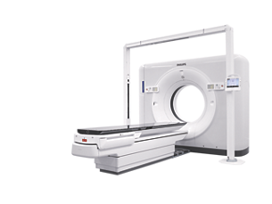
Big Bore RT is designed as a CT simulator to enhance clinical confidence, accelerate time to treat and maximize value of its investment without compromising on patient experience – four dimensions that are essential towards excellent care.
View product
-
MRCAT Brain
MRCAT Brain clinical application allows the use of MRI as the primary imaging modality for radiotherapy planning of primary and metastatic tumors in the brain without the need for CT. Detailed anatomical information for contouring and attenuation maps for dose calculations are both obtained from a single, submillimeter resolution 3D T1W mDIXON MR sequence. Artificial Intelligence (AI) is used for fast computation of continuous Hounsfield units directly on the MR console.
View product
-
MRCAT Prostate + Auto-Contouring
As a plug-in clinical application to Ingenia MR-RT, MRCAT Prostate + Auto-Contouring provides attenuation maps and automated, MR-based contours of prostate and organs at risk in as little as 20 minutes – all in a repeatable ‘one-click’ workflow.
View product
-
MRCAT Pelvis
MRCAT Pelvis lets you plan radiation therapy using MRI as a single modality solution. Within just one MR exam, MRCAT Pelvis provides excellent soft-tissue contrast for target and OAR delineation, and continuous Hounsfield units for dose calculations. MRCAT (MR for Calculating ATtenuation) data can be used for export to treatment planning systems for CT-equivalent** dose calculations. In addition, MR-based imaging enables CBCT-based positioning based on soft-tissue contrast with the look and feel of CT.
View product
- *Compared to overlay solution.
- 1. Compared to Philips scans without Compressed SENSE.
- 2. Compared to the Ingenia 1.5T ZBO magnet.
